

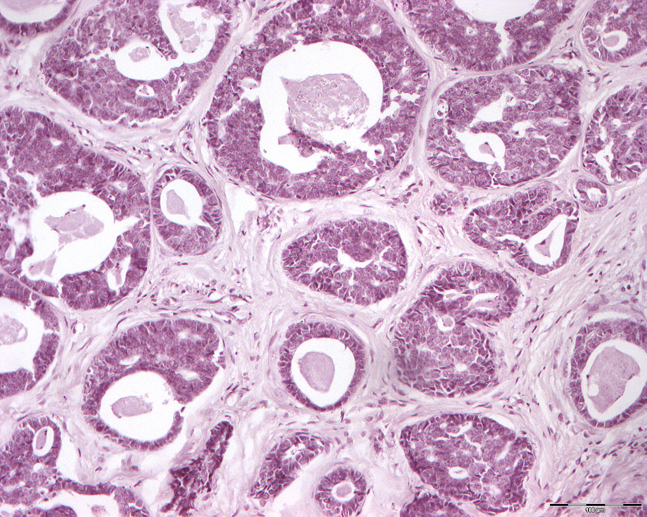
Adenocystic carcinoma (200X)
Clinical information: A 50-year-old woman had an approx. plum-sized tumor in the hard palate. The tissue was obtained from the same patient as section no. 148.
Clinical diagnosis: Metastasis from adenocystic carcinoma
Microscopic examination: Reference is made to section no. 148. Here you see the same type of tumor tissue which is poorly demarcated and grows infiltratingly. Note the tendency for growth into vein walls and into vein lumina. In one place growth is seen in the nerve sheath, which is common for malignant tumours. In one area, a certain influence of the stroma with hyalinisation is found. This is stated to be characteristic of adenocystic carcinomas.
Comment: The term cylindroma for this tumor is obsolete and refers only to the frequent occurrence of cylindrical, hyaline structures in solid epithelial masses or between solid epithelial strands. Adenocytic carcinomas with a lot of cribriform pattern are highly differentiated and have a better prognosis than cases with a lot of solid tumor tissue (little like glandular tissue). The prognosis is considered worse for adenocystic carcinomas in smaller salivary glands than larger ones.