

Adenocystic carcinoma (40X)
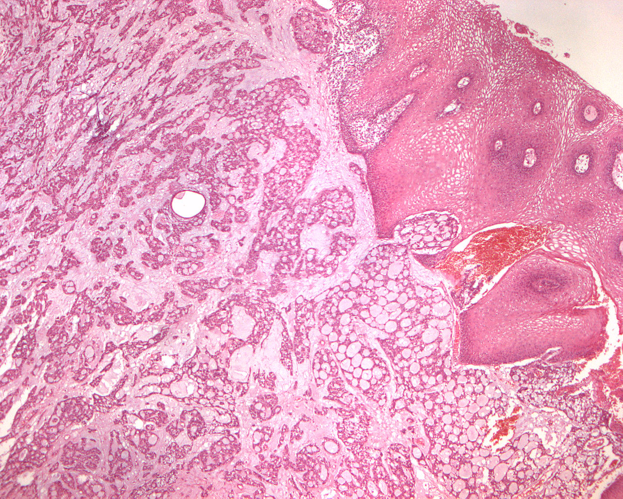
Clinical information: A 50-year-old woman had an approx. plum-sized tumor in the hard palate. The tissue was obtained from the same patient as in this section of adenocystic carcinoma.
Clinical diagnosis: Tumor palati.
Microscopic examination: The tumor tissue shows several morphological patterns; you can partly see flakes, strings and tubular structures consisting of isomorphic (iso. Greek = equal) epithelial cells interspersed with some lighter colored cells with dark nuclei. In some areas you can observe that the tumor tissue has the typical adenocystic appearance with islands of tumor of varying sizes and that the tumor tissue is perforated by small and larger holes (cribriform = sieve-like pattern; cribrum(L) = sieve). The stroma is a loose connective tissue that appears slightly mucinous in som areas. The tumor is not encapsulated and grows infiltratively in the surroundings; locally it "attacks" the epithelial surface.
Comment: Adenocystic carcinoma is a rare and aggressive form of cancer that most commonly arises in the salivary glands, but can also occur in other areas of the body, such as the breast and trachea. This malignancy is characterized by its slow but relentless growth pattern and a propensity to invade surrounding tissues and nerves. Adenocystic carcinoma is known for its unique histological features, with cells forming glandular structures and cyst-like spaces like the ones seen in this section. Despite its slow growth, it tends to metastasize to distant organs over time, making early detection and treatment crucial for improved outcomes. Treatment typically involves a combination of surgery, radiation therapy, and sometimes chemotherapy, tailored to the individual patient's specific circumstances. Due to its rarity and the challenges it poses, ongoing research is essential to develop more effective therapies for this uncommon but formidable cancer.