


Salivary gland with salivary ducts, acini, artery and veins
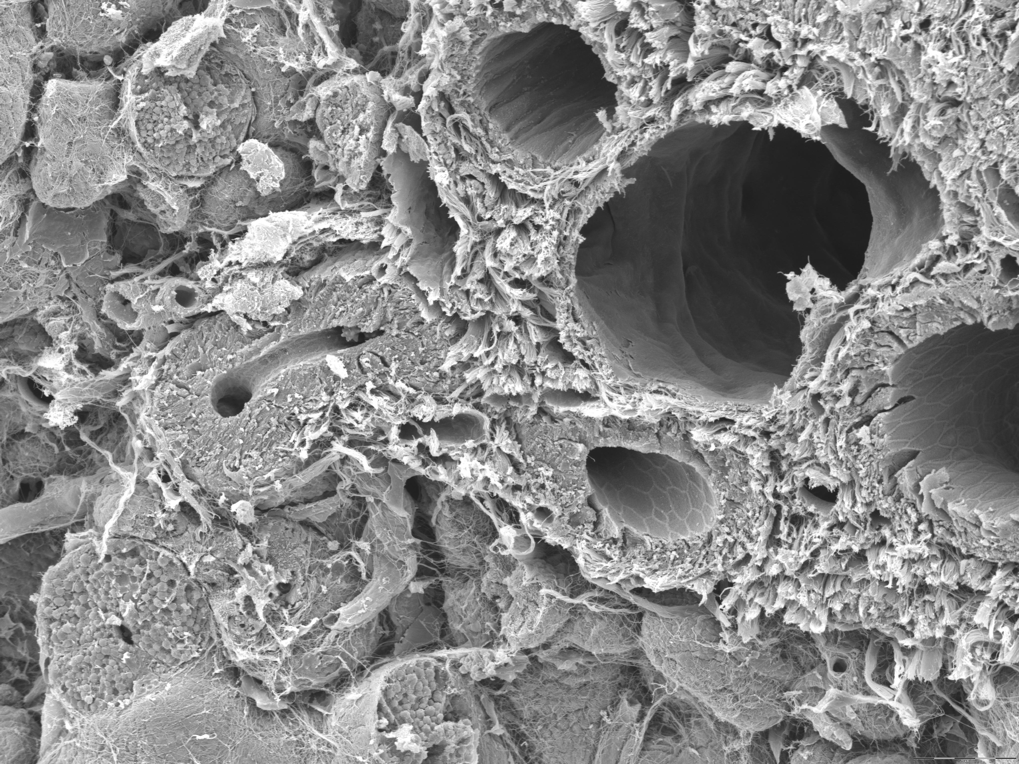
This is a scanning electron microscope image of a salivary gland. This image really show off the potential of the scanning electron microscope. The salivary gland has been cut open and it reveals many structures. The larges structure is a vein flanked by an artery and a salivary duct. The wall of the vein is thin and smooth in contrast to the artery which has a thicker wall and lined with smaller protuberances. If you take a closer look at the inside of the salivary duct you'll se a pattern. This pattern represents the borders between the epithelial cells lining the salivary duct.
Quite a few salivary acini are also visible, both cut open or with an intact surface. Secretory granules and the lumen of the acinus can also be seen.
See if you can find all these structures in this histology section of the submandibular gland.