


Artery, vein and excretory duct of the submandibular gland (200X)
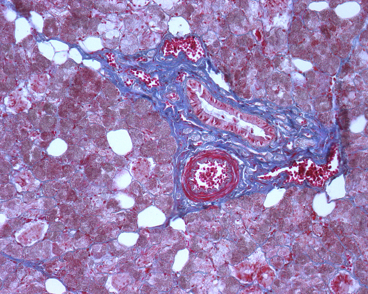
Embedded in connective tissue, a salivary excretory duct is circled by an artery and a few smaller veins. The connective tissue is surrounded by glandular tissue and a few fat cells. As mentioned, the connective that is found within the septa and in between the acini, stains blue.
The image is well suited to study the difference in appearance of arteries and veins. While the artery has a thick muscular wall, the wall of the vein is thin and contains no muscle. The arteries appear round, the veins flat. There is also a scanning electron microscopic image of a salivary gland. See if you can spot some of the same structures in that image.