


Dentine with dentinal tubules
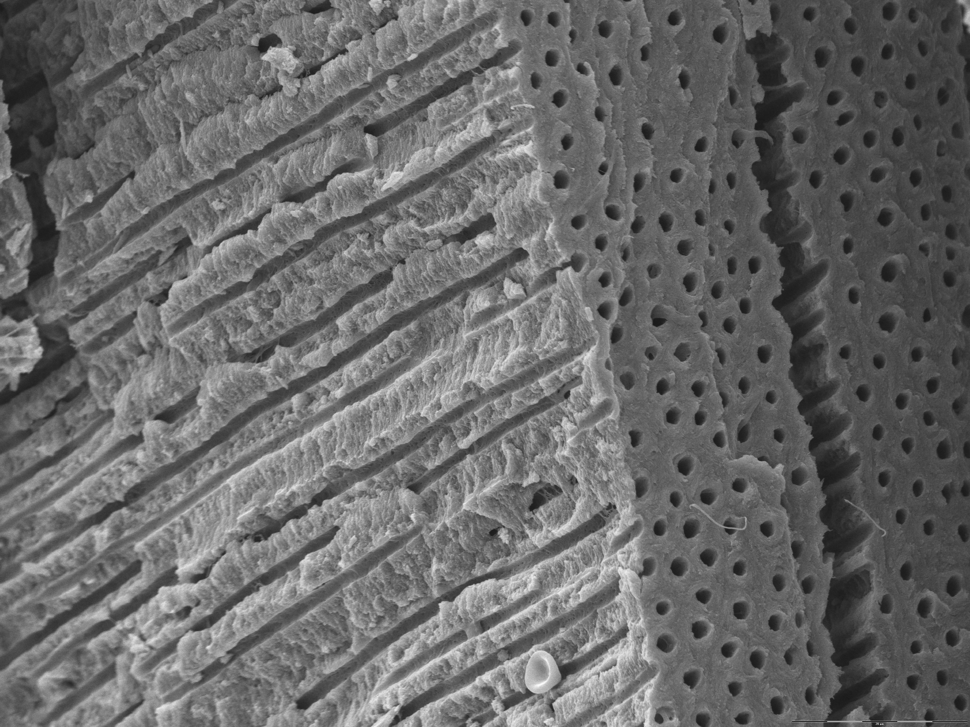
This is an image acquired using a scanning electron microscope and shows a piece of dentine. It illustrates the number of dentinal tubules and their size (size may vary as to what part of the dentine you look at and the age of the individual). An erythrocyte can be seen at the bottom of the image telling us about the scale involved (the diameter of an erythrocyte is approximately 7,5 µm). Erythrocytes are usually not seen in the dentine due to the fact that there are no blood vessels in the dentine. This erythrocyte must have originated from elsewhere, maybe some damaged soft tissue.
Compare this image to the image of dentinal tubules close to the dentino-enamel junction (DEJ).