


The periodontal sulcus (200X)
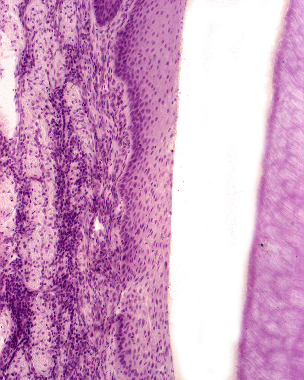
This is a magnification of the bottom of the periodontal sulcus just coronally to the enamel cementum junction. The enamel is not visible bacause it has been washed away during the praparation of the tissue sample. But it leaves behind an empty space I've chosen to call 'the enamel space'.
The connective tissue just below the epithelium displays only a few inlammatory cells. There is no sign of periodontitis indicating that the tooth might come from a young individual.