


Gingiva and periodontium (40X)
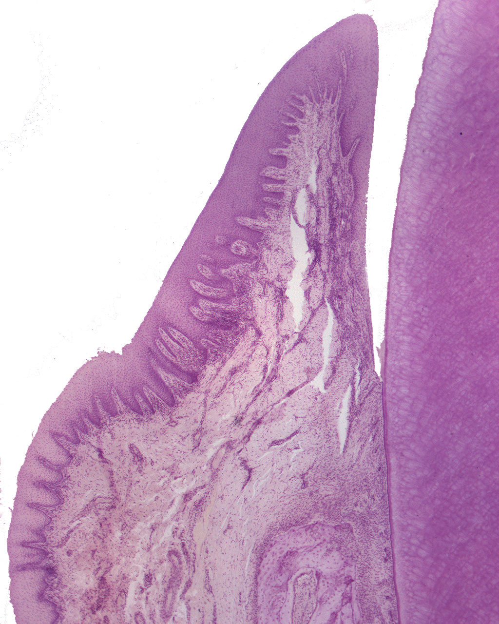
This is a section through a molar and its supportive tissues. Only the buccal surface of the tooth and its adjacent gingiva is shown in this image. Notice the gingival epithelium. Rete pegs have formed (strands of epithelium), stretching some distance into the lamina propria. The bottom of the gingival sulcus ends at the enamel cementum junction (ECJ). If you take a closer look at the connective tissue lying just below the sulcular epithelium, you will only see a few inflammatory cells. There is no sign of periodontitis, and if you think this tooth might come from a young individual, your're absolutely right. This is also evident if you look at the thickness of the cementum; it's very thin indeed.