


Demineralized tooth with inflamed gingiva (40X)
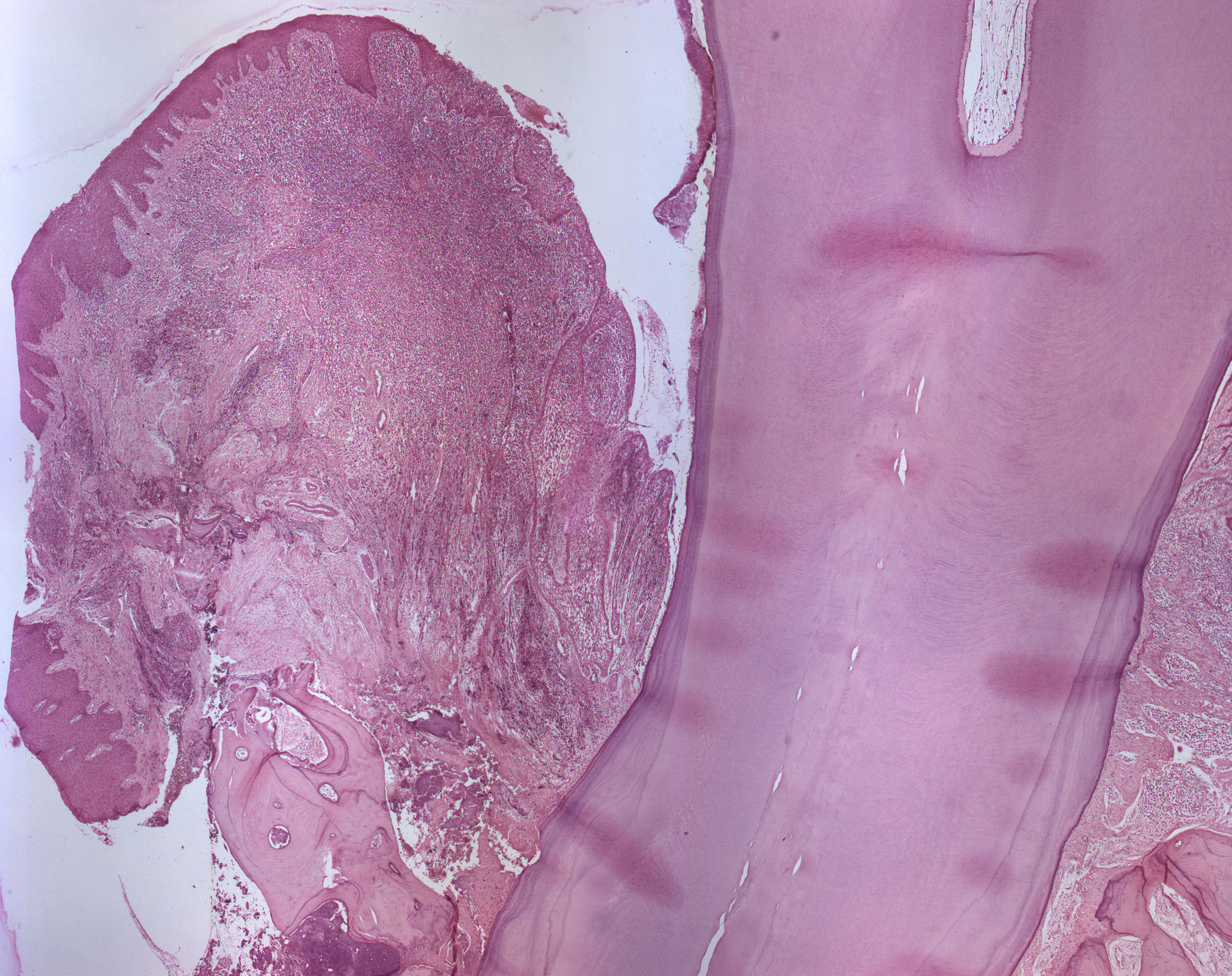
This is a longitudinal section through a piece of inflamed gingival and the adjacent tooth. Most of the supportive tissue of the tooth is lost and the root surface is covered by plaque. The periodontal ligament and bone can be seen towards the apical part of the tooth although the apex is not visible.
The cementum covering the dentine is quite thick, indicating that the tooth originates from an old individual. Incremental lines are seen throughout the cementum.