


Erupting tooth (40X)
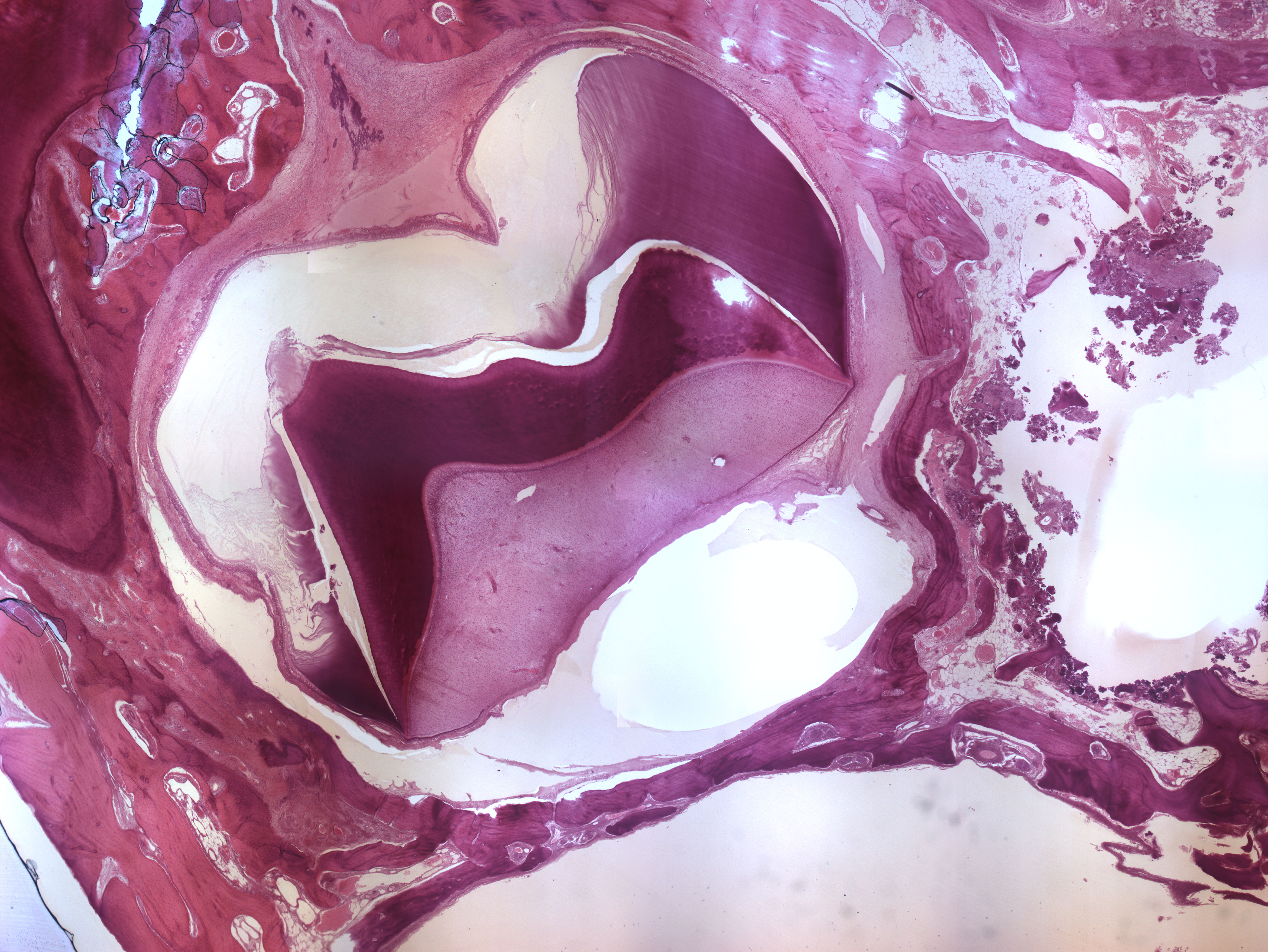
This is a section through a jaw showing a developing premolar (center) and a decidous molar (top). Most of the enamel of the premolar has disappeared during the decalcination process and left an empty space (white area of enamel - enamel space). This is not the case in the cervical area because the maturation of the enamel is not finished in this location.
There are a few white areas (cracks) in between the enamel and dentine due to the sectioning of the tissue sample. These white areas are artifacts and should be disregarded as normal anatomical structures. The predentine stains light red and can be seen as a narrow zone below the dentin. The unmineralized enamel stains violet.
At the position of the future cemento-enamel junction (CEJ), the epithelial root sheath of Hertwig can be spotted. It can be identified as a thin strand of epithelium bilaterally.