


Stria vascularis and endolymph (200X)
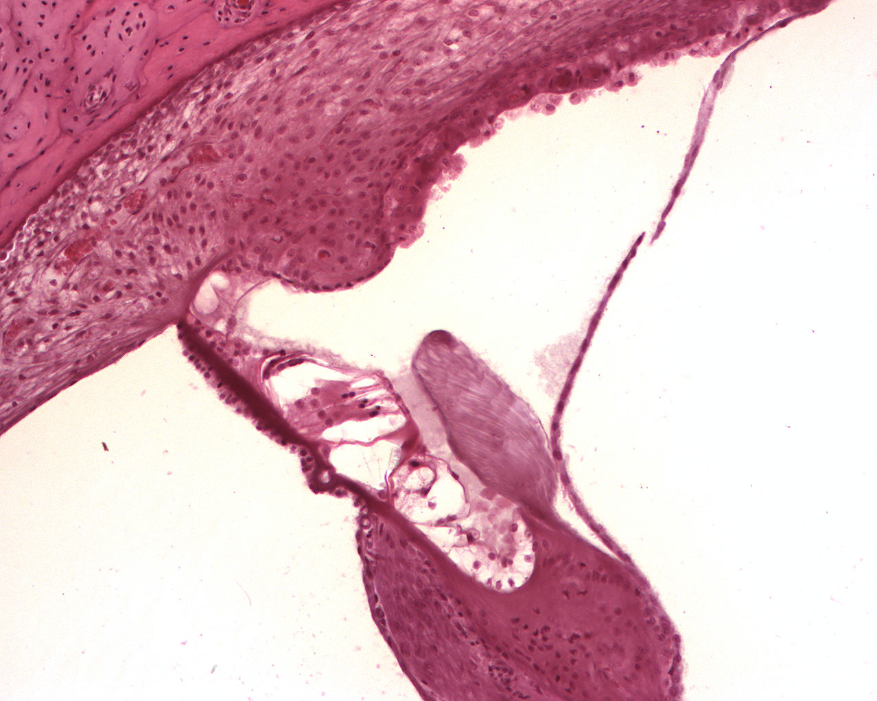
Afferent nerve fibers originating from the organ of corti can be seen at the bottom of the image, temporal bone at the top. It is agreed upon that, at least to some extent, the stria vascularis lining the bony wall of the ductus cochlearis is responsible for producing the special quality of the endolymph. It is believed that the cells of the epithelial lining contibutes to the high concentration of K+-ions in the endolymph. Due to this high concentration of K+-ions, the endolymph maintains a +80mV membrane potential against the perilyph, and the hair cells of the organ of corti develop an unusually high membrane potential against the endolyph, about 150mV.