


Cochlea and labyrinthus (40X)
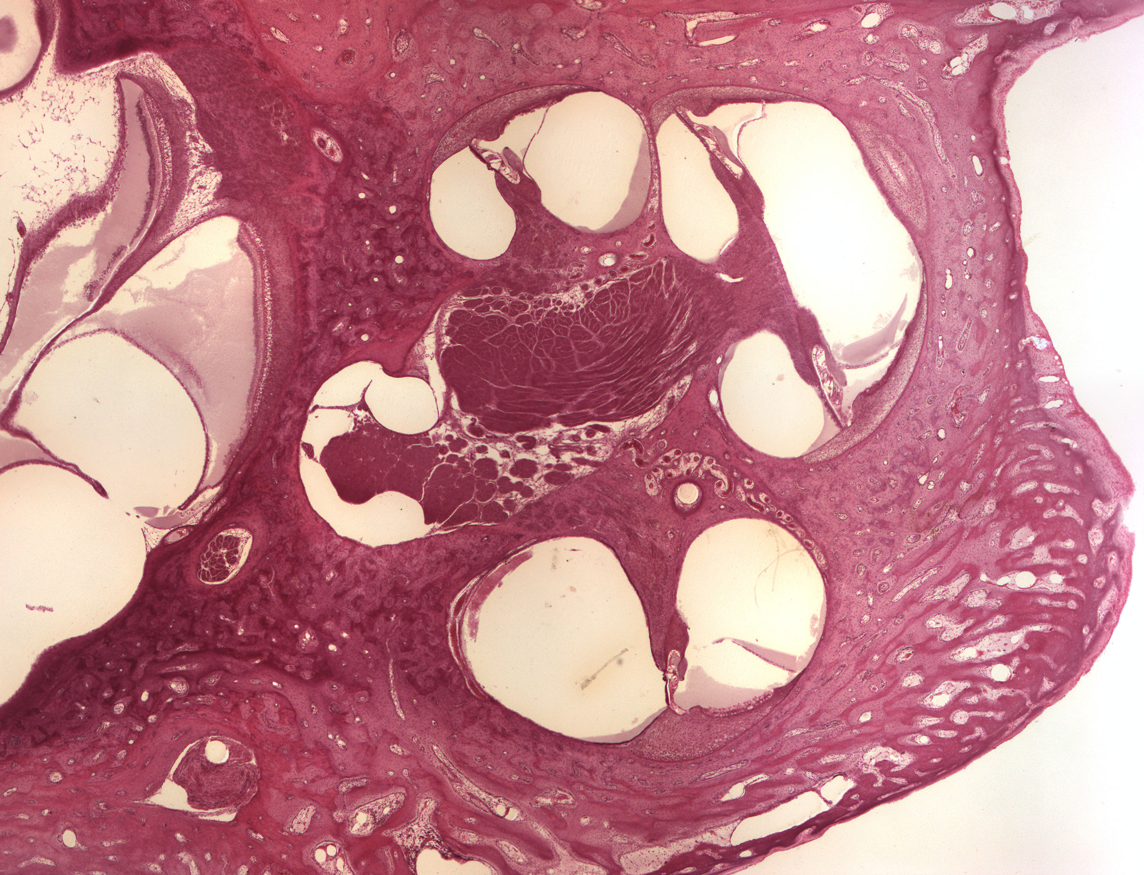
This is a section through the inner ear. You can see the "twists and turns" of the cochlea and the different spaces within: the scala vestibularis, ductus cochlearis (scala media) and scala tympani. The spiral ganglion can be seen at the center. Most of the visible tissue in this image is bone.
Some parts of the labyrinthus cochlearis can be spotted on the left hand side of the image.