


Vestibular system (40X)
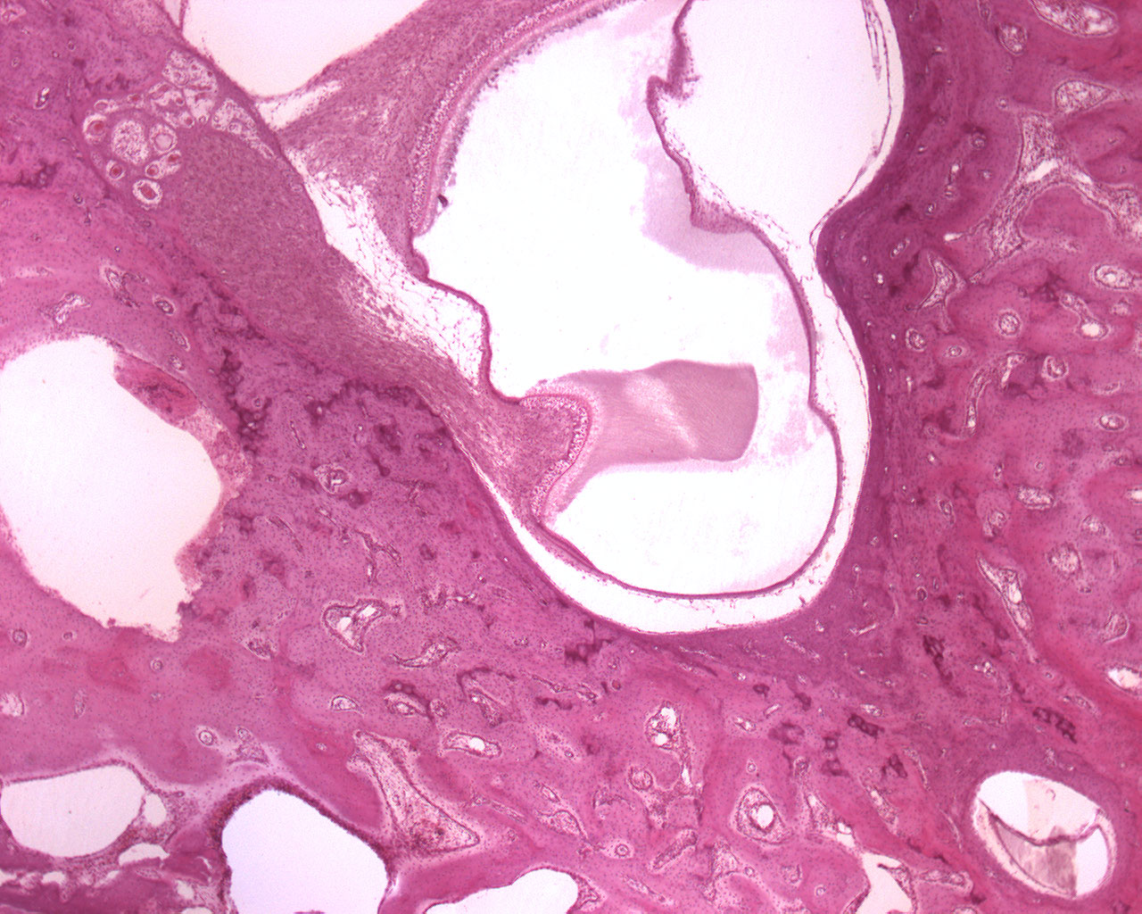
This is an image of the vestibular system showing both the bony labyrinth and the membranous labyrinth. I strongly urge you to study your anatomy atlas or follow the link at the bottom of this text to get a grip of the structures macroscopically as well. I find theese structures really confusing, so try to get the grips on the macroscopic anatomy and physiology first as it will help you in understanding these set of images.
The membranous labyrinth is a collection of fluid filled tubes and chambers which contain the receptors for the senses of equilibrium and hearing. Only the structures responsible for equilibrium is seen here. The membranous labyrinth is lodged within the bony labyrinth and has the same general form; it is, however, considerably smaller and is partly separated from the bony walls by a quantity of fluid, the perilymph. In certain places, it is fixed to the walls of the cavity.
The membranous labyrinth contains fluid called endolymph. The walls of the membranous labyrinth are lined with distributions of the acoustic nerve, also known as the vestibularcochlear nerve.
Within the osseous vestibule, the membranous labyrinth does not quite preserve the form of the bony cavity, but consists of two membranous sacs, the utricle, and the saccule.
The membranous labyrinth is also the location for the receptor cells found in the inner ear.
The macula of utricle allows a person to perceive changes in longitudinal acceleration (in horizontal directions only).
Anatomy and physiology of the inner ear: http://www.ilocis.org/documents/chpt11e.htm
Source: Wikipedia/Grays Anatomy