


Tooth bud and Mekcle's cartilage (40X)
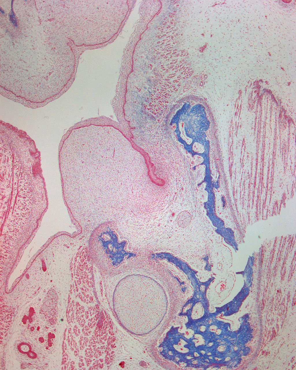
Here are a few details from the mandible. The dental lamina with its tooth bud both in the mandible is visible at the center of this image. The mesenchyme is condensated around the bud.
Just below the tooth bud, you can see the alveolar nerve (lat: n. alveolaris). Meckle's cartilage can also easily be spotted. The tongue is also developing. It consists of muscular fibers oriented in different directions. To the right of the tongue in this image, you can see glandular parenchyma.