


Head of a human fetus (7 cm)
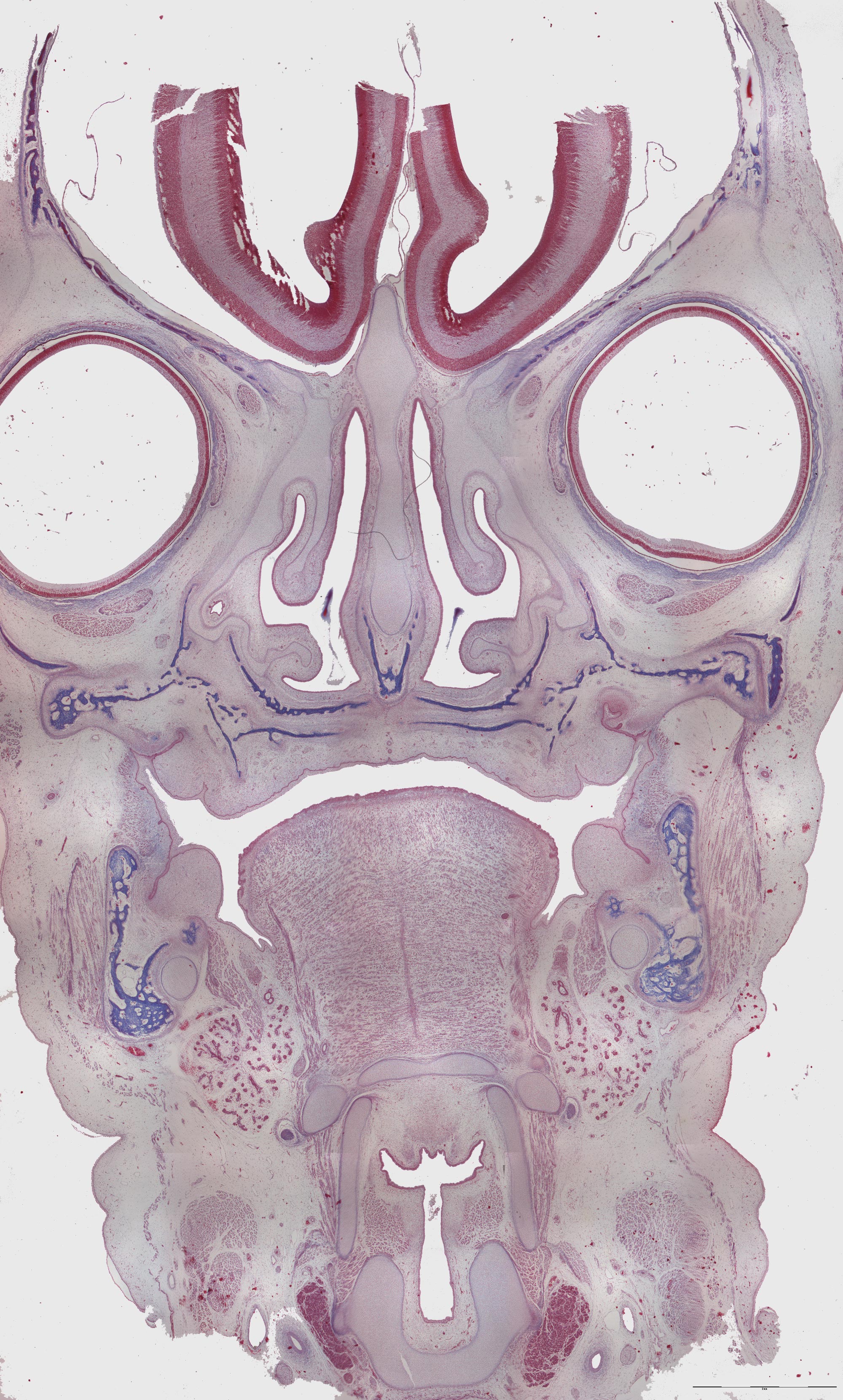
This is a frontal section of the head of a human fetus. In contrast to one of the other head sections, the tissue is well preserved showing many structures.
The nasal cavity (lat: cavum nasi) is divided in two by the nasal cartilage within the nasal septum. At both sides of the septum, you can see the nasal conchas (lat: concha nasalis media et inferior). At this stage of development the concha consist of cartilage .
The palate and the maxilla also contain a few spicules of bone. In addition you can see the dental lamina with its tooth bud both in the maxilla and the mandible. The mesenchyme is condensated around the bud.
Just below the tooth bud in the mandible, you can see the alveolar nerve (lat: n. alveolaris). Meckle's cartilage can also easily be spotted. The tongue is also developing. It consists of muscular fibers oriented in different directions. At both sides of the tongue, you can see glandular parenchyma. Cartilage comprising parts of the larynx can be seen below the tongue.