


All layers of the retina (200X)
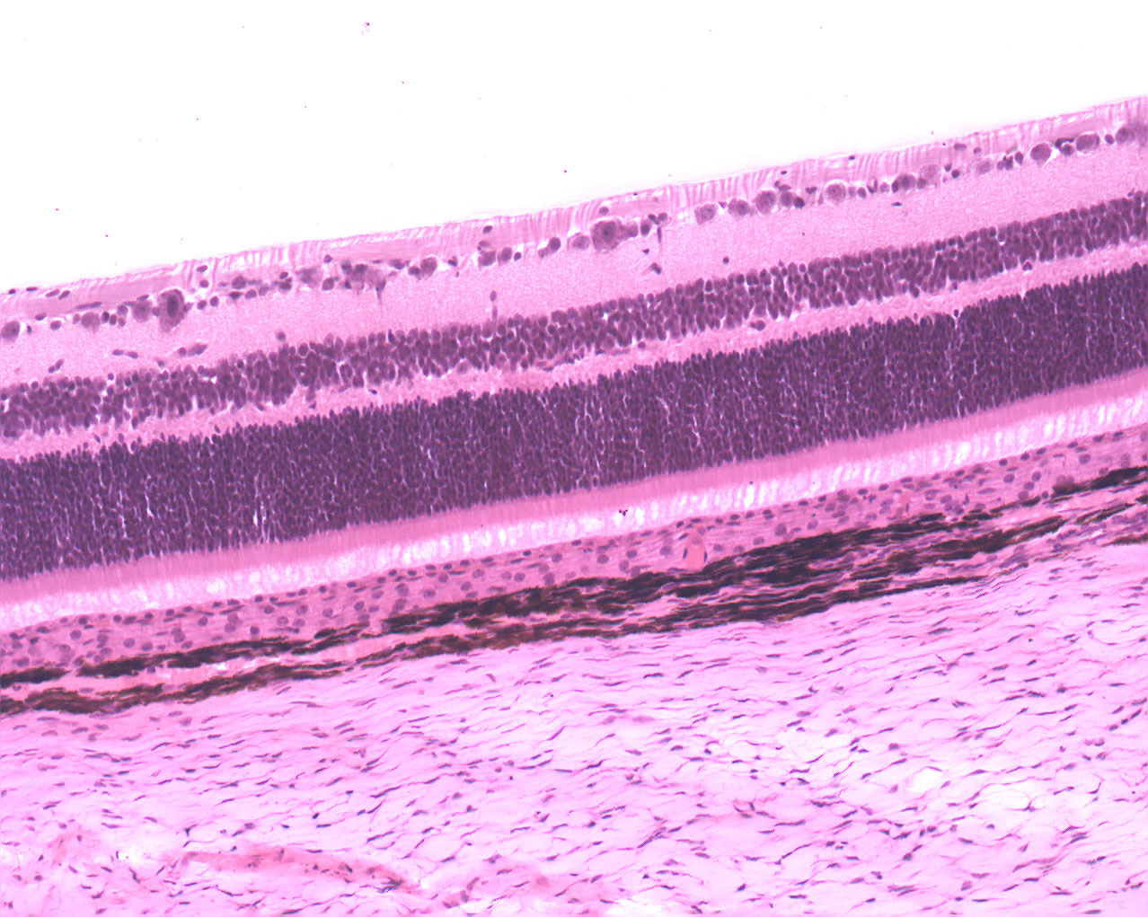
This image shows all layers beautifully. Just to make some order of the mess, I've made a short list of all the layers starting from the outside of the eye (bottom of image) to the inside (top). I find it quite easy to get confused by all the names. That's why I made a list:
- Most peripherally lies the sclera which consists of fibroblasts, a few elastic fibers and bundles of collagen of which all run parallel to the surface of the eye.
- Choroid is the dark membrane above the sclera.
- Pigmented epithelial cells; dark and thin layer (not visible in this image).
- Stratum photosensorum or the receptor layer is a thin layer that stains lightly. This is the rods and cones.
- Outer nuclear layer holds the nuclei if the rod and cone cells.
- Outer plexiform layer is a synaptic layer.
- Inner nuclear layer holds Müller's cells and nuclei of bipolar nerve cells.
- Inner plexiform layer is a pure synaptic layer with synapses between the bipolar cells and the ganglon cells.
- Ganglion layer: gangon cells
- Optic fiber layer conists of Müller's fibers, including horizontal and vertical axons from ganglion cells (lightly stained layer).
In between the stratum photosensorium is a unknown layer of cells containing distinct nuclei.