


Principal fibers, resorbtion and supportive tissues (40X)
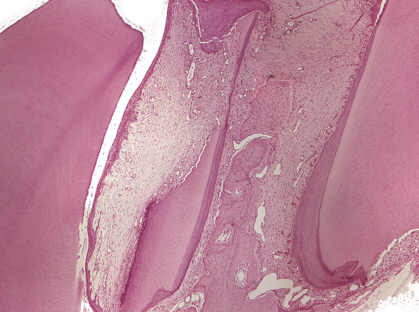
In some regions of the section, the principal fibers (periodontal ligament), their orientation and attatchment to the alveolar bone can be seen.
The cementum stains darker than the dentine and thickens the closer to the apex you look.
The apex of the deciduous tooth (the tooth to the right) is resorbed. There is also a small root positioned in between the two teeth. If you take a closer look, you'll see a few resorbtion lacunae and odontoclasts.