


Shedding of teeth (40X)
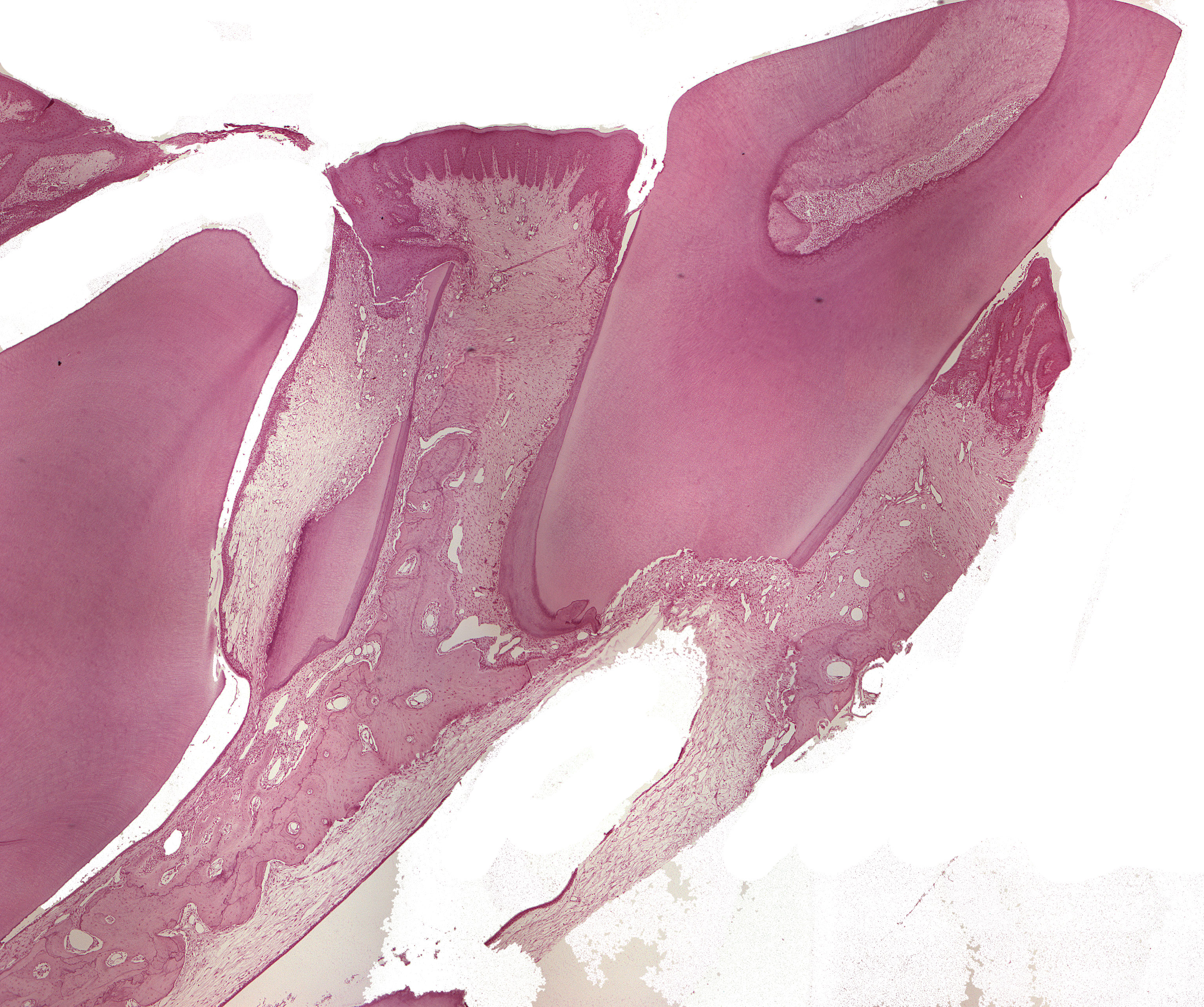
This is a section through a jaw of a monkey showing a deciduous tooth and a developing permanent tooth including their supportive structures. The gingiva surrounding the deciduous tooth is keratinized. There is a small inflammatory reaction in the tissue beneath the sulcular epithelium, but it is hard so spot in these set of pictures. The bottom of the gingival pocket is seen some distance apically to the enamel cementum junction along the cementum surface. This indicates that some of the tooth's supportive tissue has been broken down.
The apical area of the deciduous tooth is in the process of being resorbed. Coronally, tertiary dentine has formed, probably due to attrition.
If you look closely, there's even a third root visible in between the deciduous tooth and the permanent tooth. All is left is some dentine and cementum. The alveolar bone is seen in the middle of the image.
The root of the permanent tooth is still open apically (not possible to see in this picture), but it near its completion and ready for eruption. The reduced dental epithelium has fused with the oral epithelium.