


Endochondral ossification (200X)
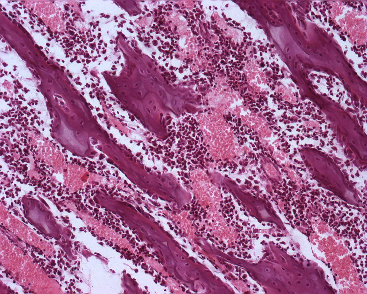
This is a detail from the diaphysis.The growth of the bone has matured. You can see quite a few spicules of bone and bone marrow. In some places, there are pieces of bluish cartilage inbetween the bony tissue. In addition, see if you can spot osteocytes, osteoblasts and osteoclasts.