


Endochondral ossification (40X)
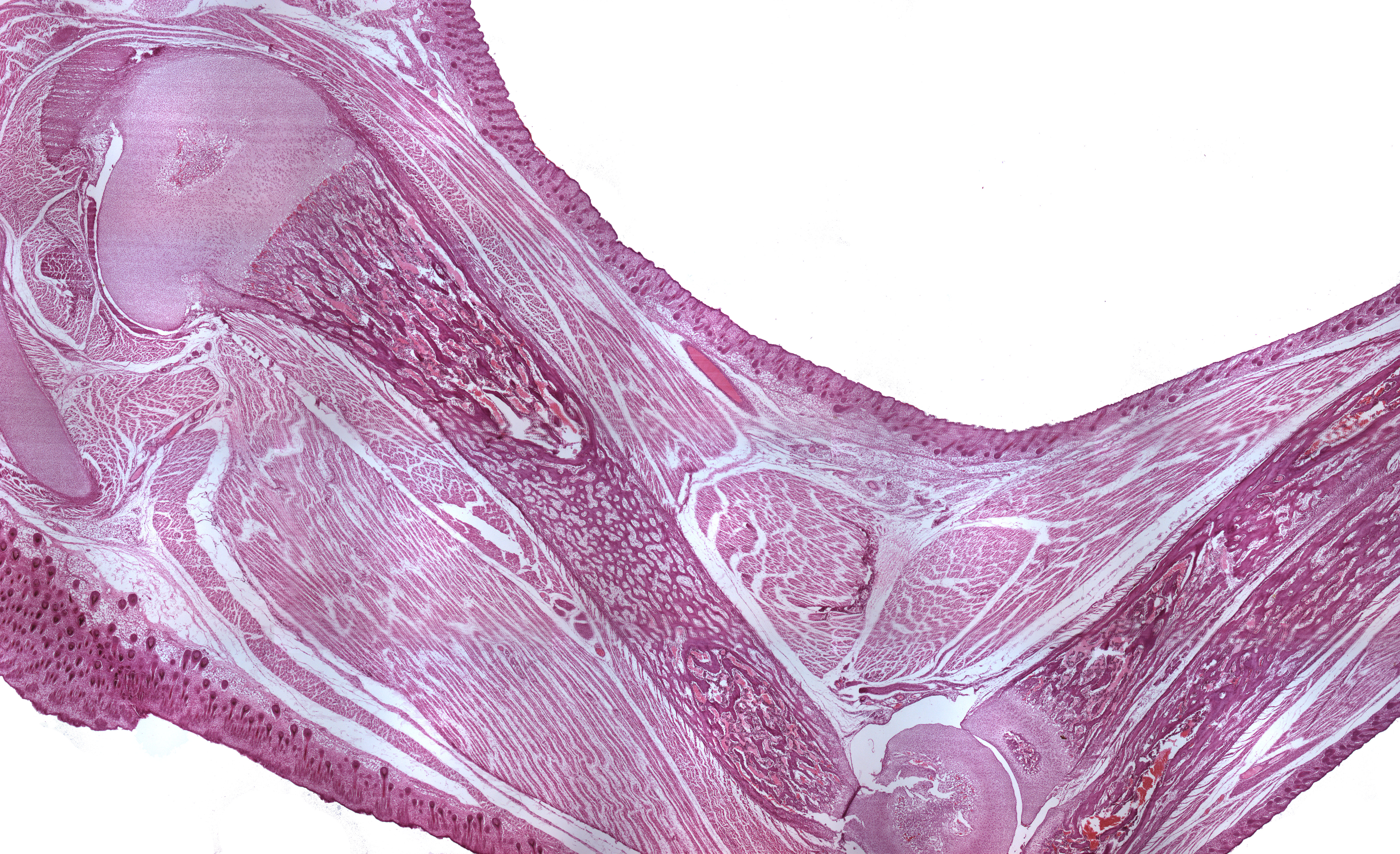
This section is taken from the leg of a guinea pig. You can see a couple of developing long bones. The surface of the specimen is covered by stratified squamous epithelium. Interspersed in the epithelium you can find hair follicles. Besides the long bones and the epithelium, you can also find muscle cells, connective tissue and blood vessels.
Because of the way the tissue is sectioned, one epifysis is seen at both ends of the diaphysis (the diaphysis is the name of the middle part of the long bone).