


Endochondral ossification (100X)
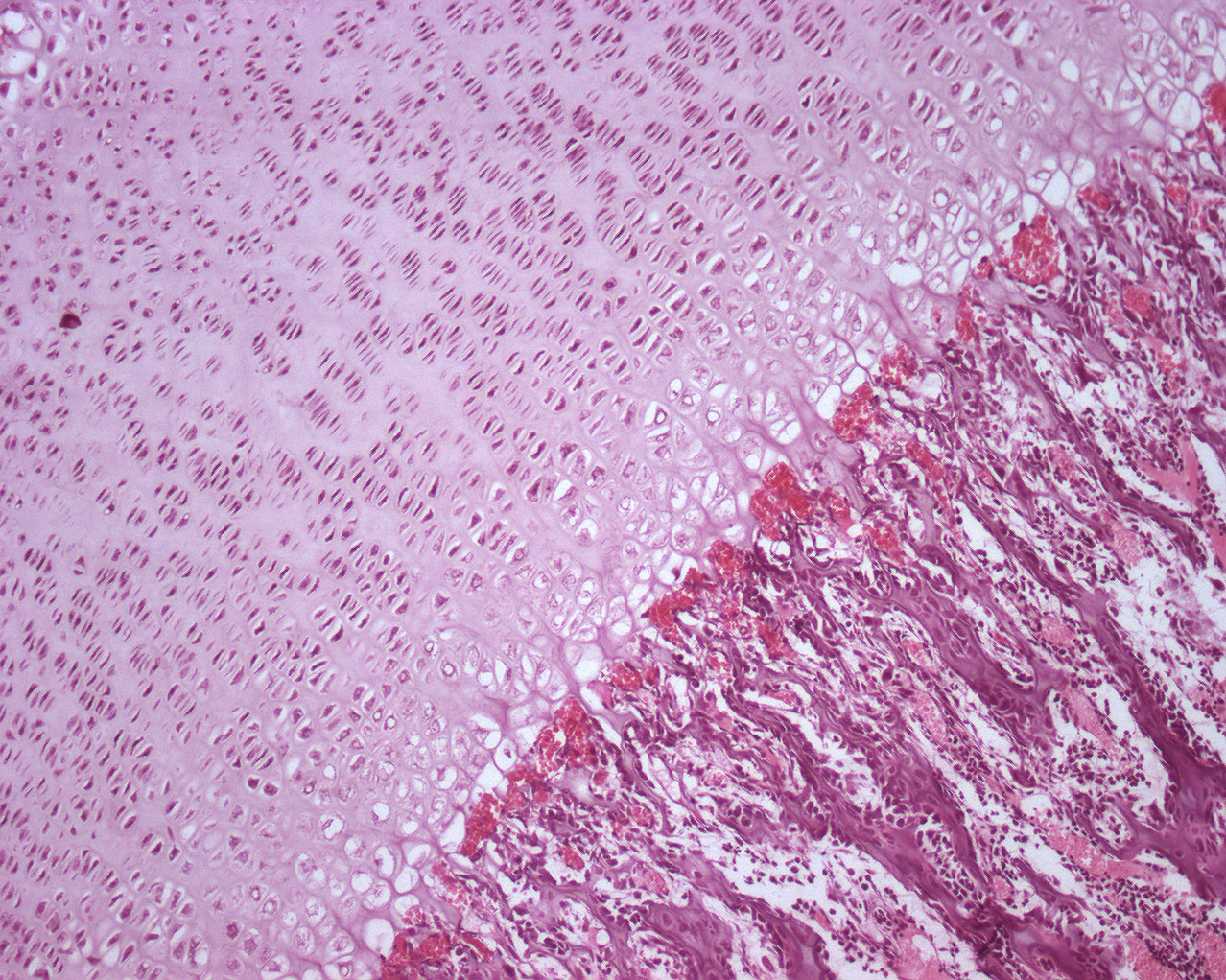
Large numbers of chondrocyes are embedded in the cartilage. The epiphyseal disc is identified by all the chondrocytes stacked in columns (not labeled in this image). Within the epiphysial disk, the chondrocytes that is closest to the epiphysis are flat, while the cells closer to the diaphysis increase in size, become cubical and undergo necrosis.