


Endochondral ossification (100X)
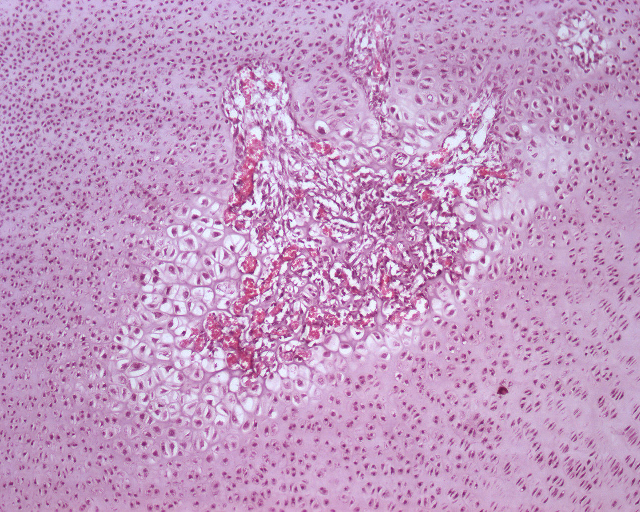
This secondary ossification center is seen in the epiphysis. A whole lot of chondrocytes is seen as well. As in the primary ossification center, the chondrocytes situated closest to the secondary ossification center increase in size and takes on a cubical shape the closer they are to the ossification center.