


Eye lid (40X)
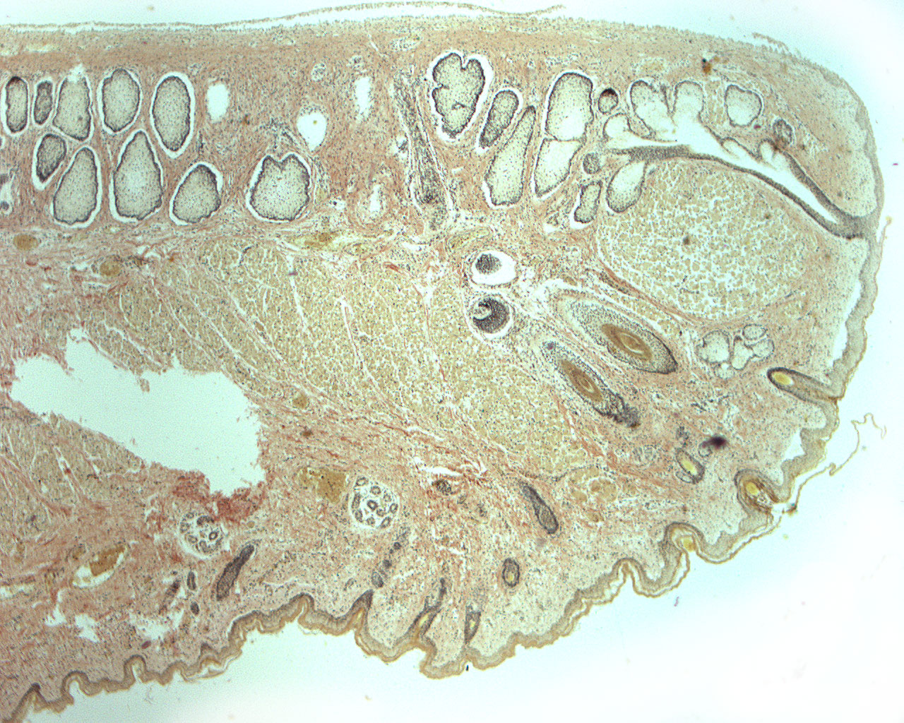
This section is showing an eye lid (lat: palpebrae). The coloring method used is the van Gieseon-stain. The outside of the eye lid is covered by the skin (stratified squamous epithelium). The epithelium facing the eyeball is of the low, stratified columnar type. Hair follicles can be seen at the anterior surface (in this image; at the bottom). These hair follicles belong to the eyelashes. Some of the eye lashes are cut in this section and color dark yellow/green. Some sweat glands are seen at the lower surface (the surface covered by skin). Try to compare this section to the H+E stained section of an eye lid.