


Soft palate (40X)
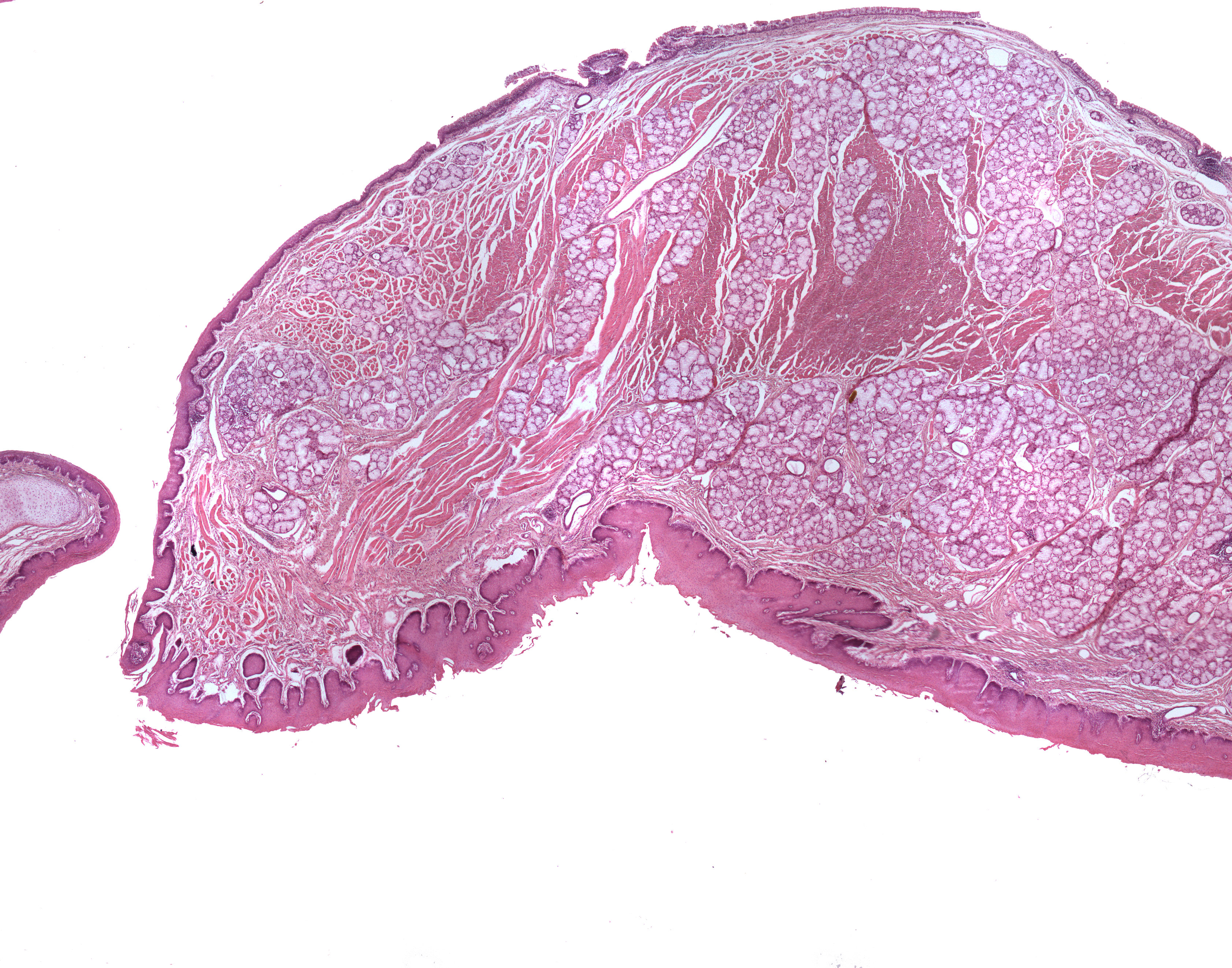
This section is taken from the pharynx of a monkey displaying the soft palate. You can see the tip of the epiglottis to the left and the soft palate to the right. In vivo, these structures lie far apart. What I've focused on in these set of pictures are the differences of epithelial lining on the oral side of the soft palate versus the nasal side.
The oral side the soft palate is covered by stratified squamous, nonkeratinized epithelium. On the nasal side, this epithelium changes character and now consists of pseudostratified ciliated columnar epithelium with goblet cells.