


Submandibular gland (40X)
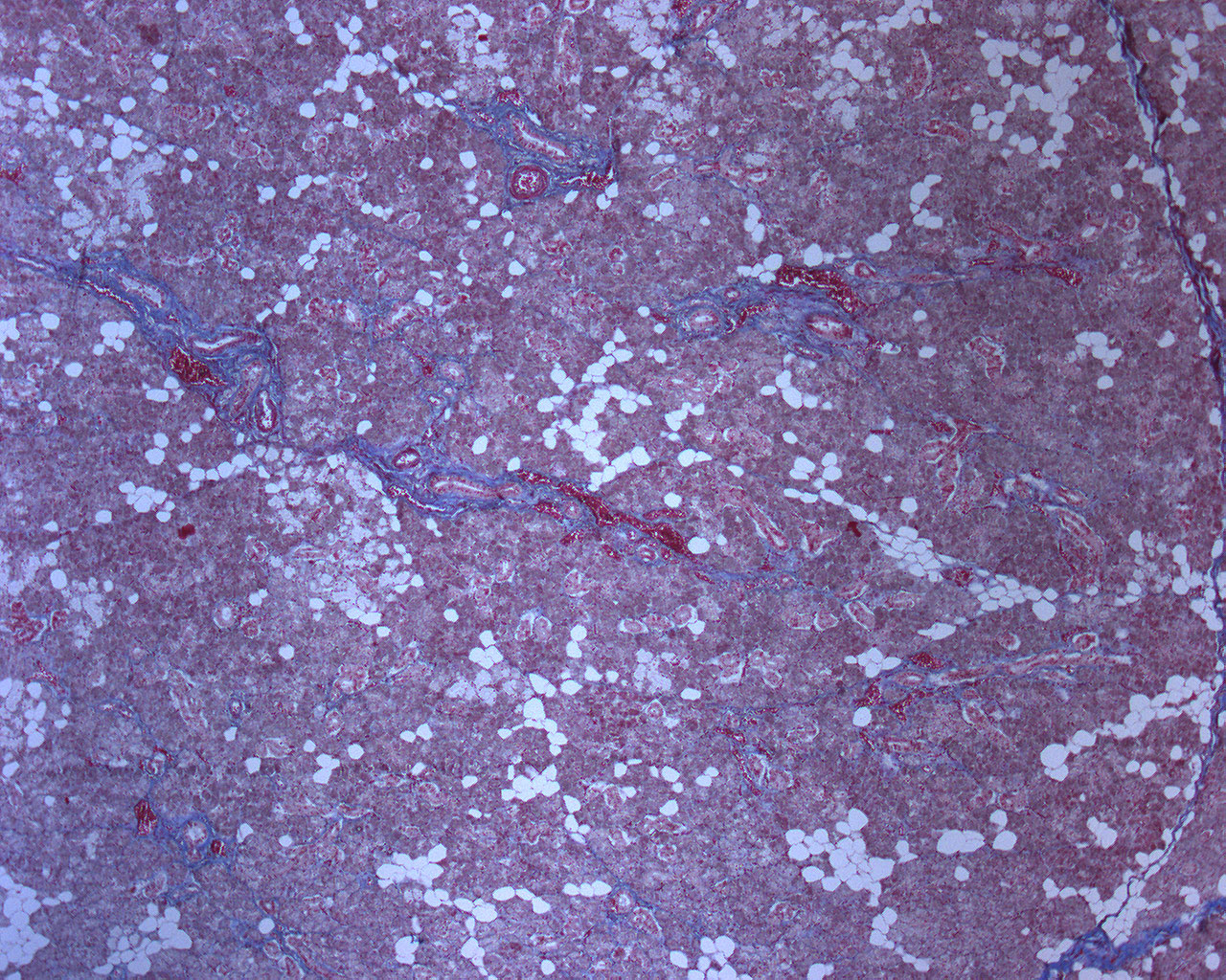
This azan-colored section is from the submandibular gland. The method used to colour the tissue sample makes the connective tissue look blue. The connective tissue spreads out between the acini in larger septa. The erythrocytes stain red and make the many vascular beds stand out. The nuclei of smooth muscle cells and myoepithelial cells also stain red. You can find the aforementioned cells in arteries and the encircling acini. The fat cells don't stain at all, except its nucleus, and leave "white patches" throughout the tissue.
The submandibular gland is mainly a serous gland, but it contains some mucous glandular tissue as well.