


Advanced bell stage with hard tissue generation (40X)
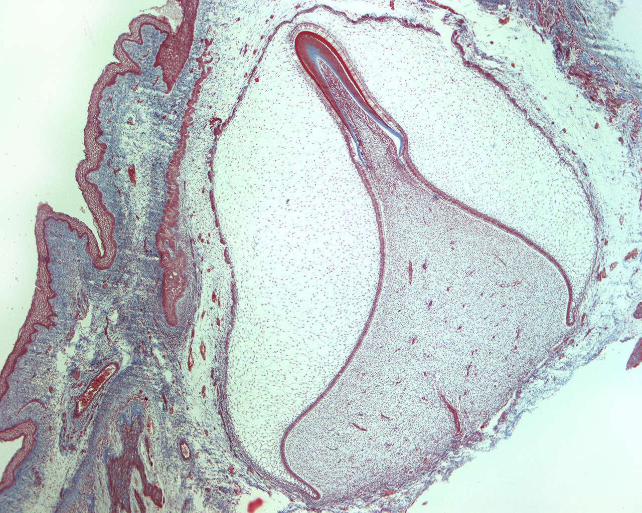
In this section, the tissue originates from the head of a human fetus and the section is made from the alveolar process. It shows a longitudinal section through the developing tooth at the advanced bell stage. The mineralization has just started at the incisal part of the tooth bud. The section is well suited to study the different layers of the tooth germ.
Some of the structures that can be seen are the lamina propria, the inner- and outer dental epithelium, the stellate reticulum and the dental papilla. Both enamel and predentine can be seen at the tip of the papilla.