
Small arteriole and venule
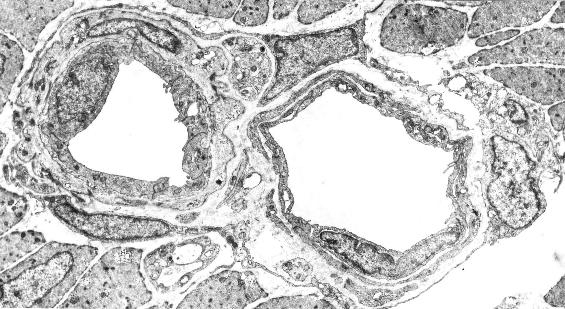
The image shows a cross-section of a very small ("terminal") arteriole and a venule. The arteriole is contracted, the endothelium is therefore tall. There is no distinct lamina elastica interna. The media consists of a layer of smooth muscle cells. Some fibroblasts are seen outside the media. The wall of the venule is significantly thinner and in some places consists only of the endothelium. Upwards between the two vessels, a small bundle of unmyelinated nerve fibers is seen.