
Continuous capillaries
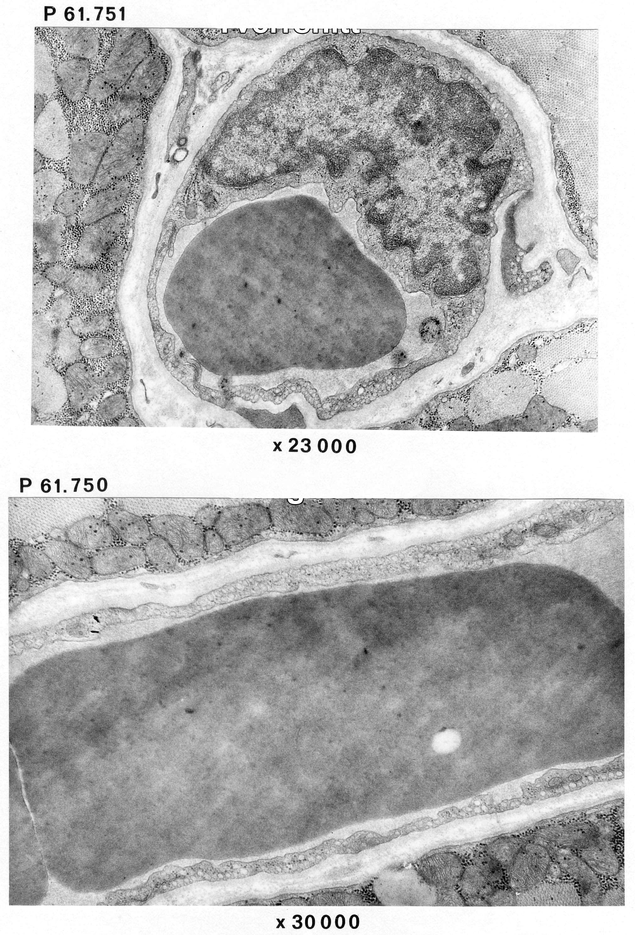
The images (P 61.751 and P61.750) show skeletal muscle from humans.
They show transverse (top) and longitudinal (bottom) sections of capillaries of the continuous type. You can also find an abundancy of endocytotic vesicles. Specialized cell contacts (cell junctions or junctional complexes) where the endothelial cells meet are seen at bottom left in the image of the transverse section (top image)
The basal lamina is thicker than in fenestrated capillaries.
Parts of pericytes are seen to the right and at the bottom of the top picture. These cells are surrounded by basement membrane both on the inside and outside.