
Discontinuous capillaries in a cat liver
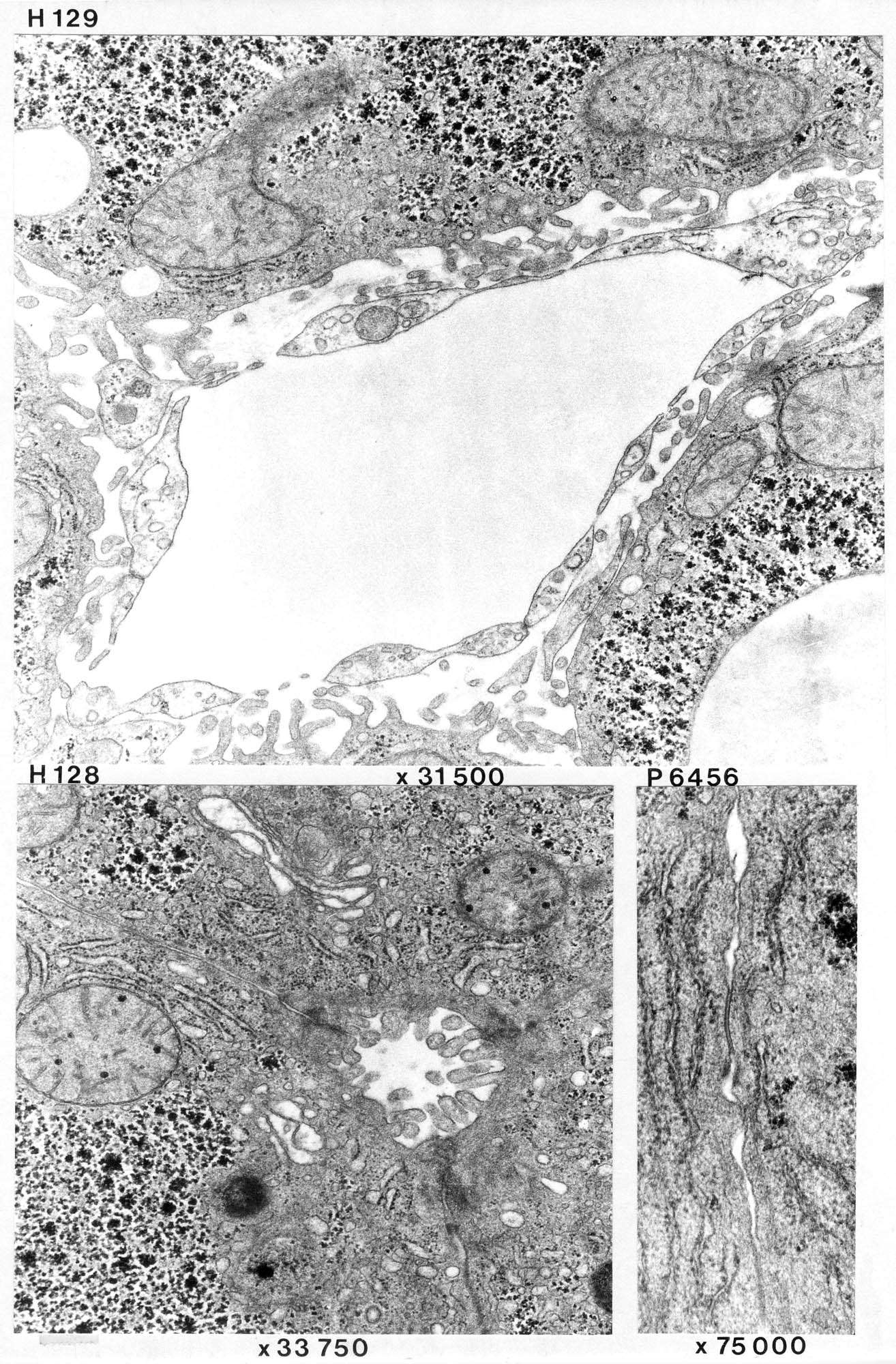
The section is from the liver of a cat.
The topmost image (H 129) shows a sinusoid with endothelial lining and the perisinusoidal space (space of Disse - the connective tissue between the endothelium and the hepatocytes). The endothelium is partly fenestrated, partly there appear to be larger openings without a diaphragm. No basal lamina. Microvilli are seen on the surface of hepatocytes and few collagenous fibrils in the perisinusoidal space.
H128 shows a bile capillary. Cell contacts between the liver cells can be seen, probably of the occludens-type near the bile capillary.
A Golgi apparatus is seen at the top of the image, otherwise abundant glycogen core and some granulated ER.
P6456 shows a specialized contact between two liver cells that is seen relatively often. Occludens type? Nexus?
Many cells in tissues are linked to one another and to the extracellular matrix at specialized contact sites called cell junctions. Cell junctions fall into three functional classes: occluding junctions, anchoring junctions, and communicating junctions.