
The proximal tubulus of the kidney from a cat
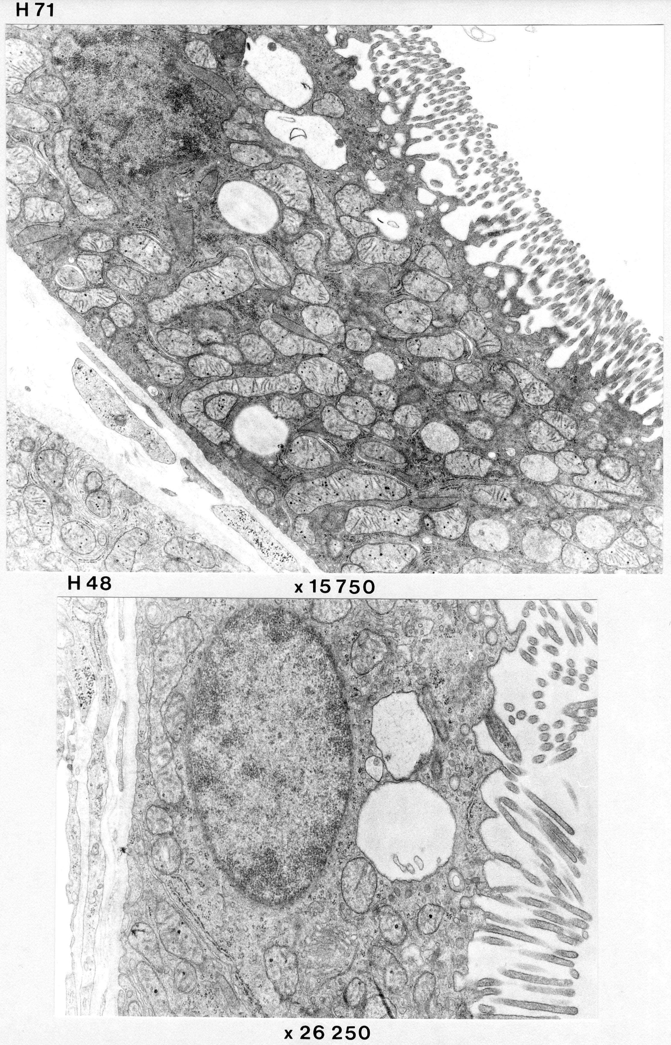
H71 shows parts of the proximal tubules with abundant microvilli and large amounts of elongated mitochondria. Basal folds are abundant and extend far into the cells, but are difficult to follow in these set of images. Also note apical vacuoles, (evidence of high transport activity?).
H48 is probably from the distal part of a proximal tubule where the epithelium is thinner.