
Glomerulus (kidney)
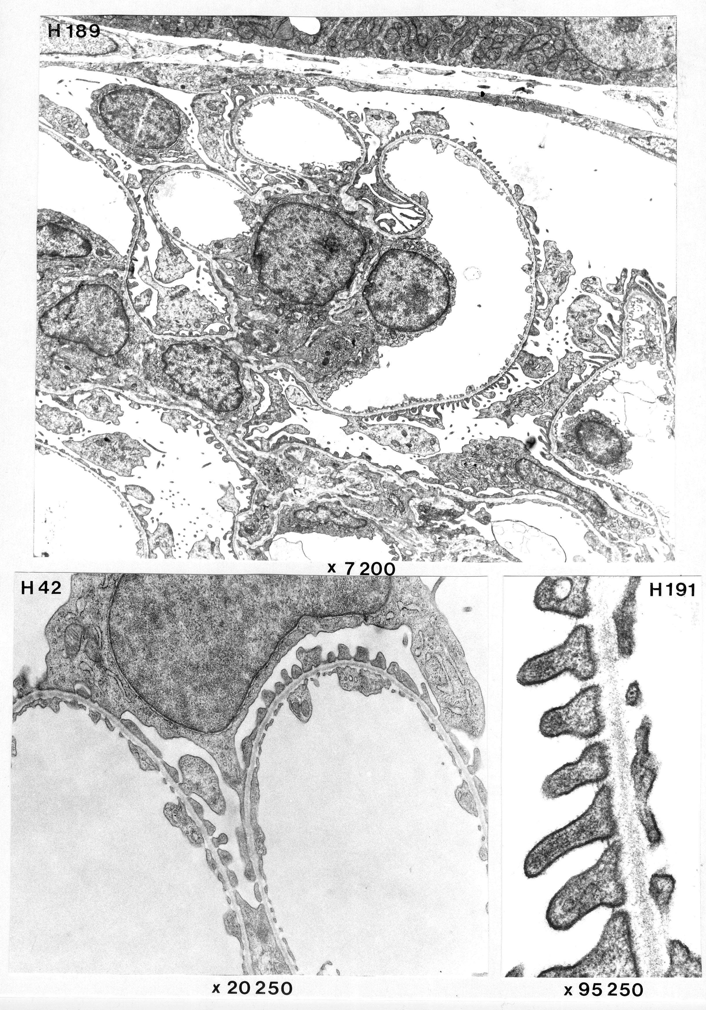
The topmost image (H189) contains parts of a glomerulus with Bowman's capsule and basal parts of proximal tubules. Podocytes, endothelial cells and 3 mesangial cells are seen.
Image H42 shows with greater magnification a podocyte and its relation to 2 capillary profiles. Fenestrated endothelium and a distinct basal lamina are also seen.
H191 shows a section of the blood-urine barrier with high magnification: podocyte feet processes on the left side and fenestrated endothelium on the right with a basement membrane inbetween. There is also a somewhat denser material between the podocyte feet processes that may resemble the diaphragm in nuclear pores.
Only a few of the fenestrae in the endothelial cells of the glomerulus have diaphragms (unlike endothelial cells elsewhere in the kidney).