
The distal tubulus of the kidney
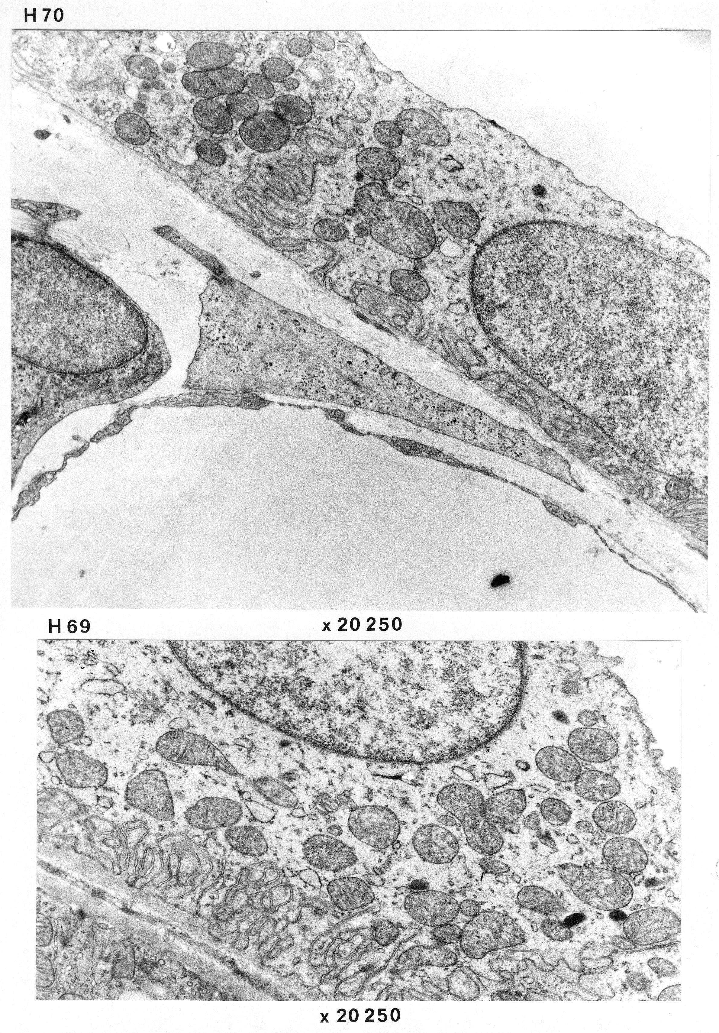
This pair of photographs (H70 and 69) of the kidney of a cat show the wall of the distal tubules. There is a lack of microvilli, abundant basal folds and folds where cells lie closely next to eachother. Notice the high degree of convultion of the membranes indicating the the surdaces of the cells must be quite interwined. In addition, you can also see a fenestrated capillary, basal lamina and some collagen fibrils.