
Macrophage
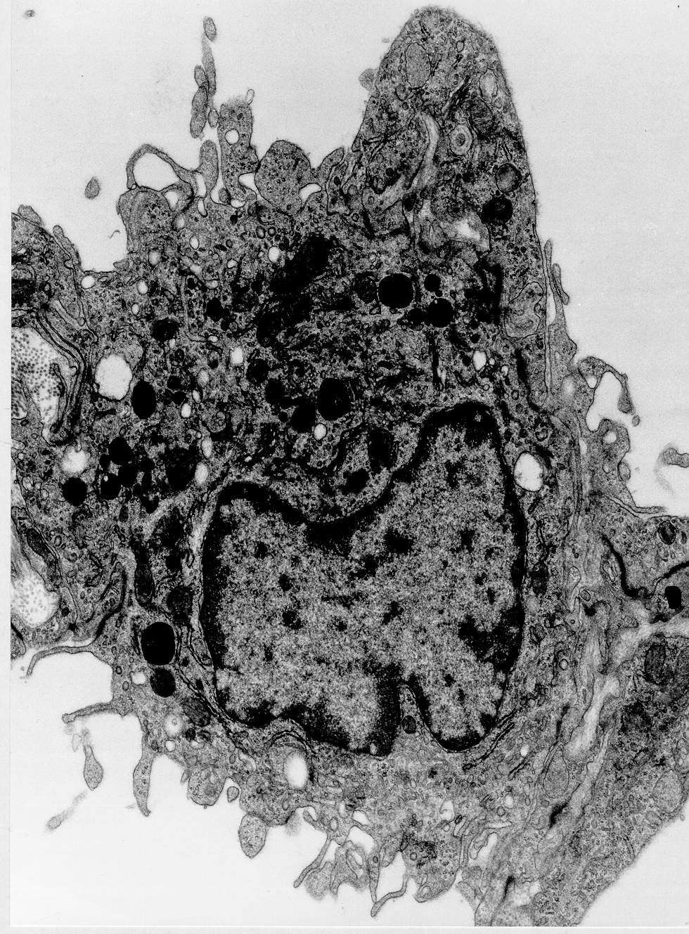
This is a typical macrophage found in a lymph node sinus. The irregular nucleus contains substantially extended chromatin. The Cytoplasm is rich in organelles: mitochondria, many Golgi complexes, large lysosomes, small flat RER sacs and numerous small vesicles and granules. Note the irregular demarcation. Do not be fooled. It is not not microvilli but thin "chambers" on the surface of the cell. Remember that yhis is a wo dimensional image of a three dimensional structure. The macrophage is seen adjacent to lymphatic endothelial cells on each side and has sent a projection through the lymphatic endothelium into collagen bundles on one side. Try to find the demarcation of the macrophage against the lymphatic endothelial cells. Junctions between the cells are probably just artefacts due to oblique cuts throuch the membranes that lie close together.