
Ischiadic nerve histology (20X)
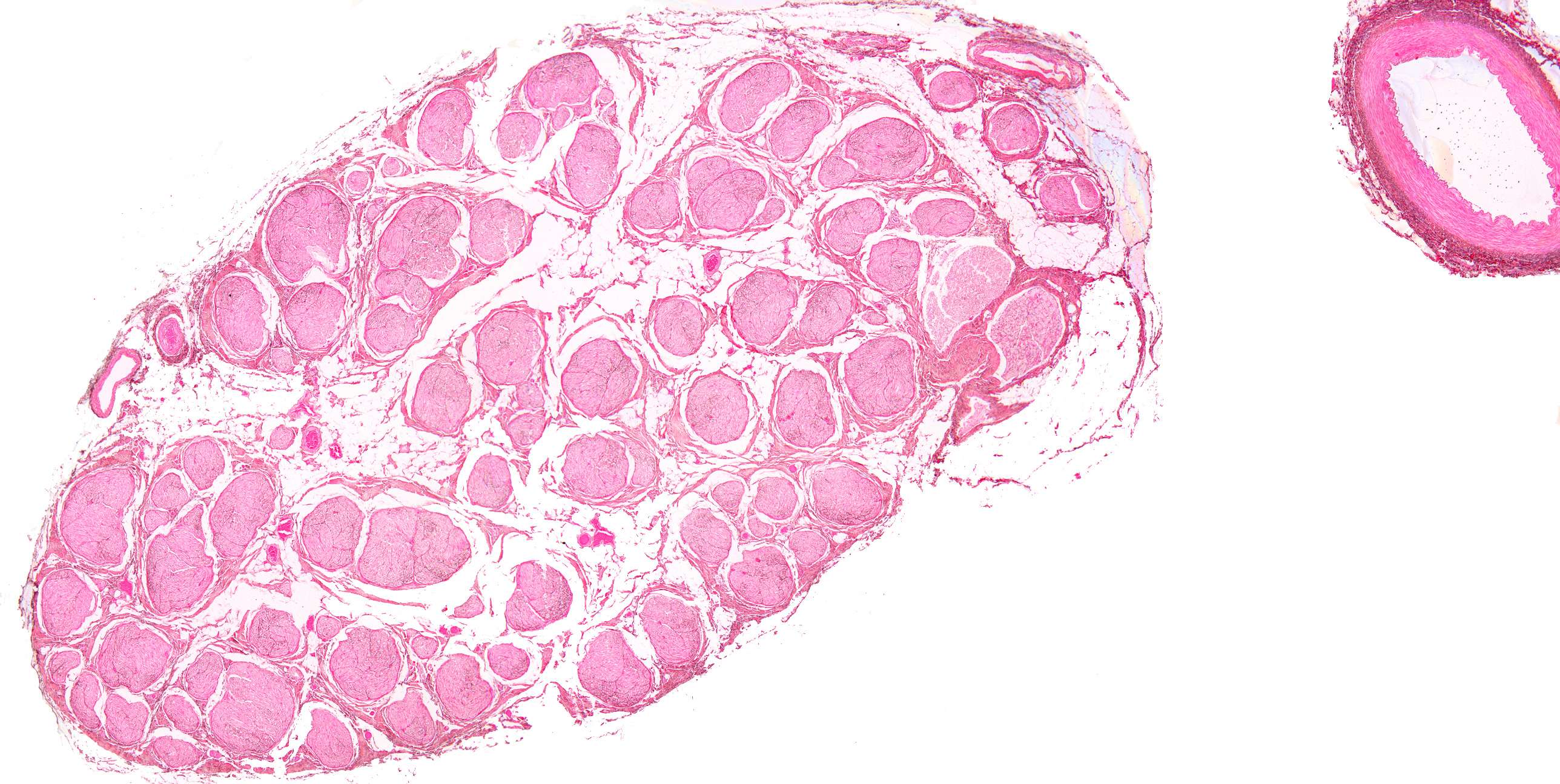
This is image a relatively low power magnification of ithe ischiadic nerve (see the range finder in the lower right corner of the image). The section consists of two structures.
Can you se the two structures?
If you guessed an artery and nerve you're correct. Distinguishing arteries from veins is usually easy as arteries have thick layers of muscle in their walls, veins do not.
Nerve fibers are divided into the following three groups:
- thick myelinated fibers
- thin myelinated fibers
- unmyelinated fibers.
The unmyelinated fibers are the thinnest. Afferent and efferent fibers are found within all three groups. The main part of the preparation contains many fascicles. Each fascicle is wrapped in a thin layer of epithelial-like connective tissue cells. It is called the perineurium and represents a permeability barrier.