
Lymph node and a high endothelial venule
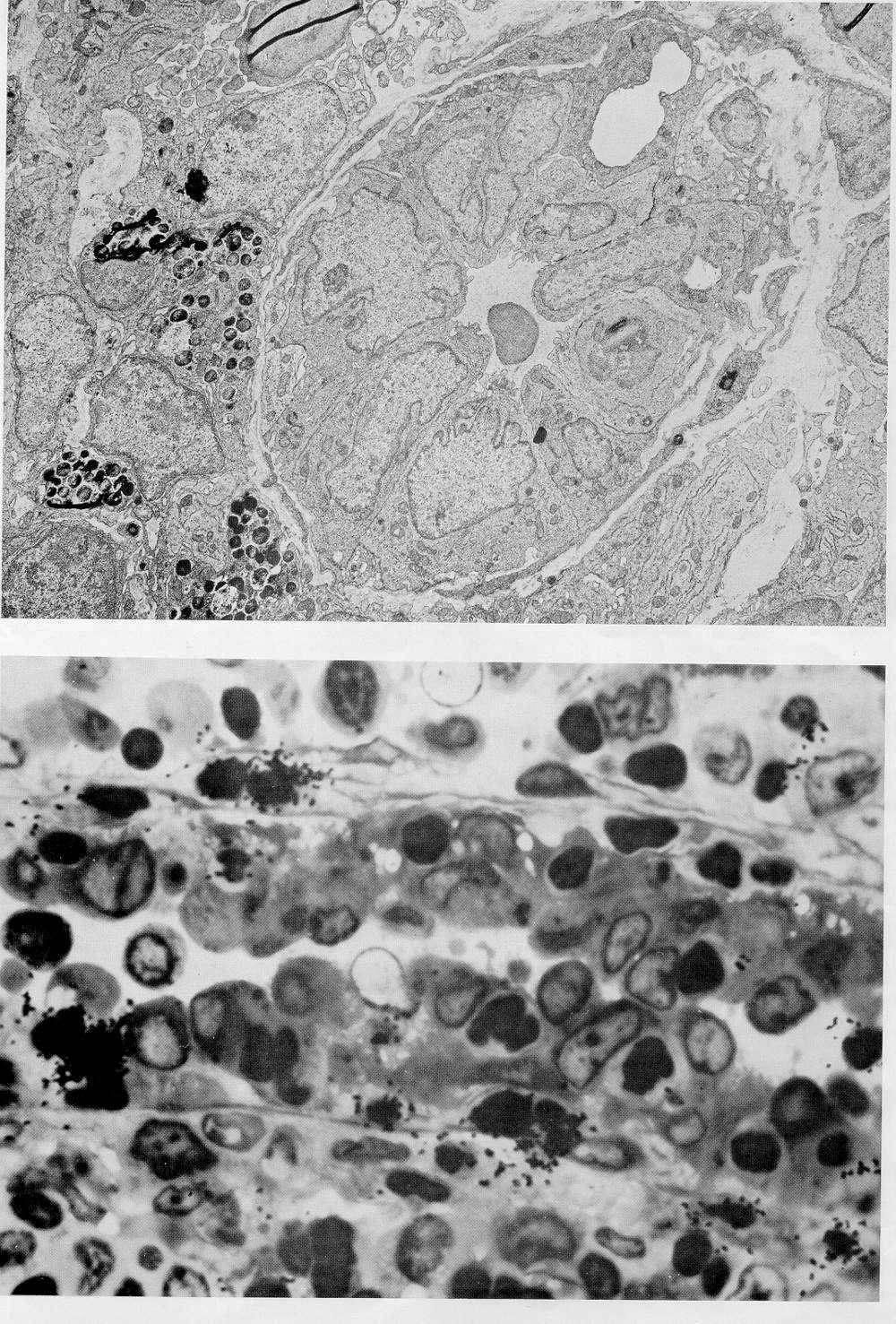
Top image: EM image of a lymph node. Note the tall endothelial cells and the narrow lumen, as well as the basement membrane and thin layers of pericytes with the basement membrane on each side. A lymphocyte is in the process of penetrating the wall to the right.
To the left, just on the outside of the vessel, there are 3 mast cells and some fibroblasts.
Bottom image: this is a light microscopic image of high endothelial venule. Lymphocytes are collected by drainage of the thoracic duct overnight. The cells are incubated with 3H-uridine and given intravenously. After 10 minutes, the lymph node is removed and localization of lymphocytes is demonstrated by autoradiography. We see here a high endothelial venule with tall endothelial cells (light nuclei) and many lymphocytes (dark nuclei). Above several of the lymphocytes that were injected into the bloodstream 10 minutes earlier, dark grains are seen that identify the lymphocytes that were given intravenously 10 min. earlier. Note that some of these cells have already penetrated the basal lamina of the endothelium and are outside the paracortex.