
Pulmonary alveolus and pneumocytes from a cat
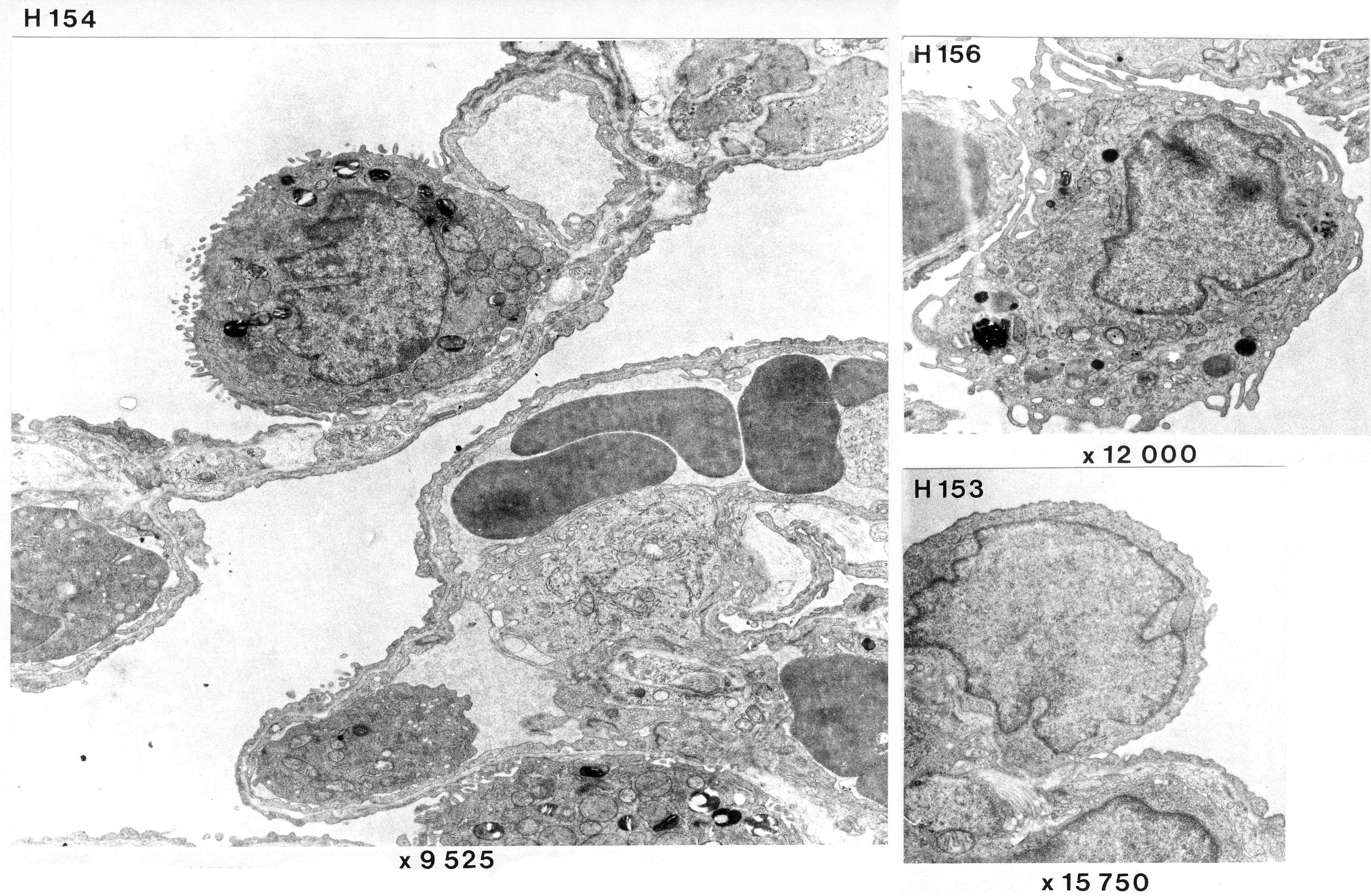
H 154: the topmost cell is a secretory cell (pneumocyte type II) which most likely produces surfactant. Note that it is embedded in the alveolar epithelium. Characteristically for this cell, intracellular lamellar secretory granules are visible. These graules stain strongly with paraphenylenediamine, which may fit with the fact that they contain phospholipid. Sparse microvilli are seen on its surface. At the bottom of the picture you can see some parts of another secretory cell.
H 156 shows a typical alveolar macrophage. This is located "on top" of the alveolar epithelium.
H 153 demonstrate the cell nucleus of an alveolar squamous cell (pneumocyte type I). These neuclei are rarely seen (much rarer than secretory cells) as a result of the fact that each cell covers a large area.