
The blood–air barrier of the lung
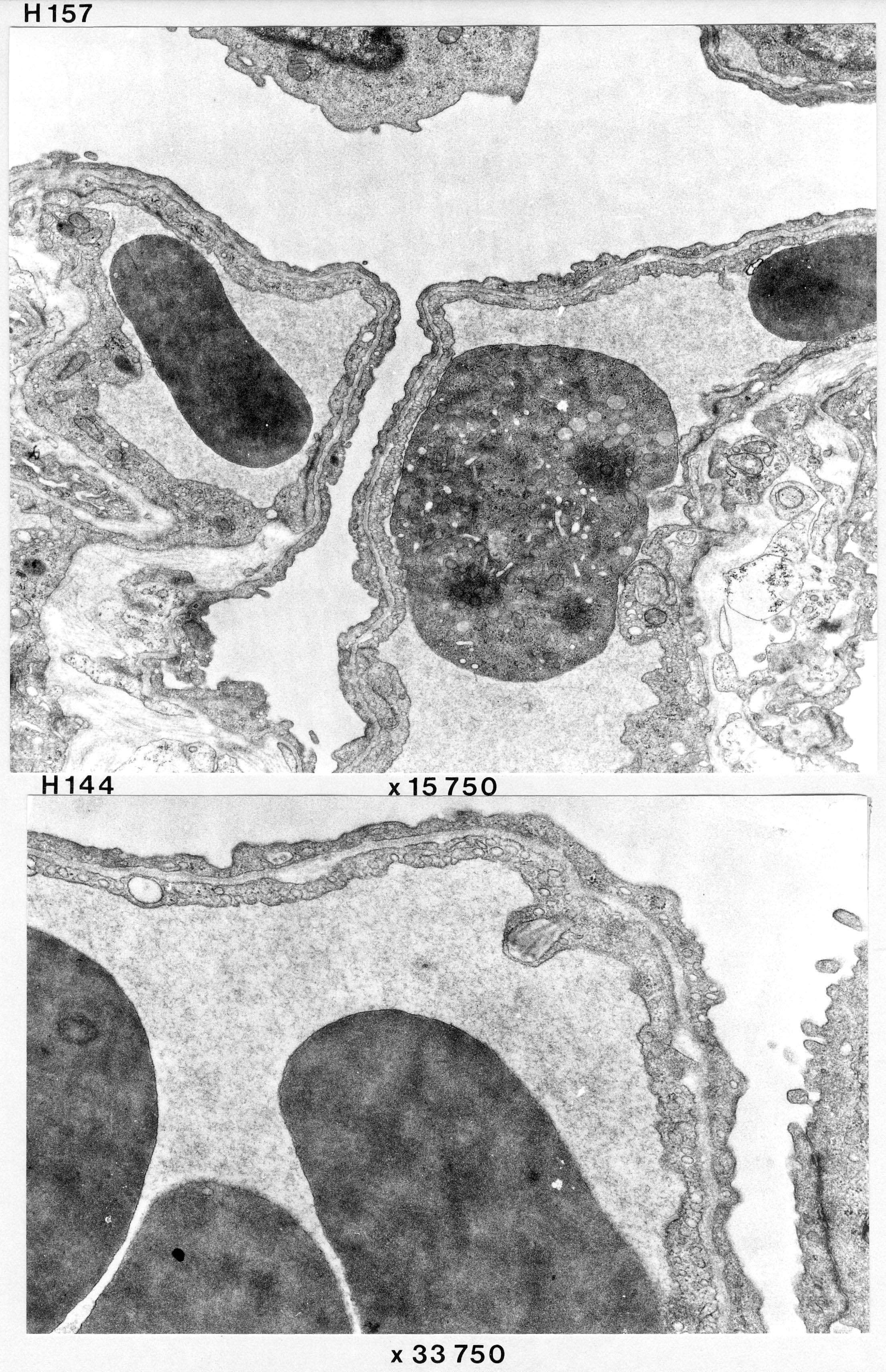
We see the layers of the alveolar wall. Blood cells are seen in the capillaries. The alveolar wall itself consists of two layers of epithelium, one from the endothelium and one from the pneumocyte, and a relatively thin basal lamina between these two types of cells. Actually there are two thin laminae: one for alveolar cells and one for endothelial cells, so the arrow in the images are actually pointing to the space between the two laminae. In some places, collagenous fibrils and thin offshoots from fibroblasts are seen.