
Tracheal epithelium
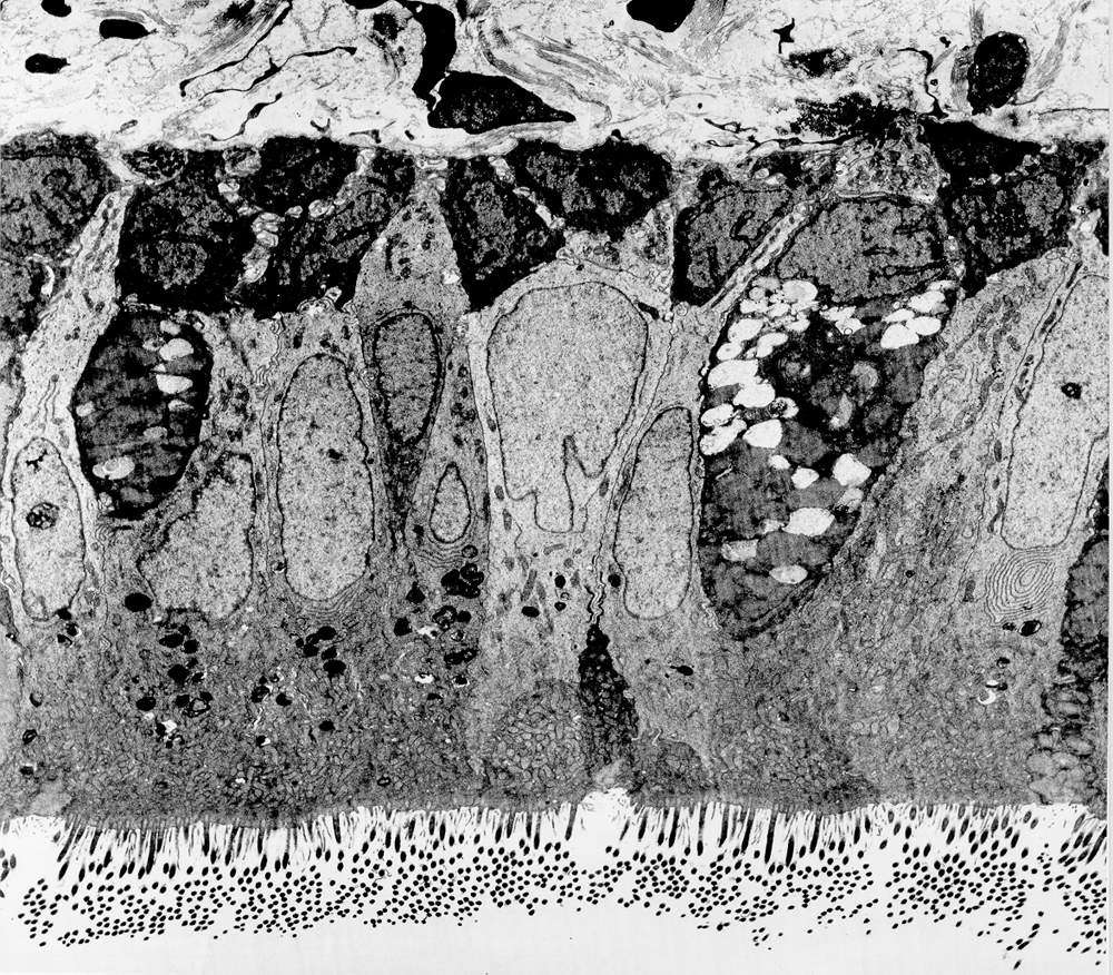
This section is from the trachea of a cat and is magnified about 5500 times(!). Next to the cylindrical epithelial cells in the middle of the image, two goblet cells can be seen (only some parts of the cells are visible). There are also cilia on the surface of the epithelial cells. Note the inside of the cells how the mitochondria are accumulated apically just below the cilia.
The basement membrane under the epithelium can barely be seen as a thin line. Two mitochondria-filled profiles are also seen situated completely basally, which probably represent nerve terminals (cf. Cough reflex).