
Trachea (20X)
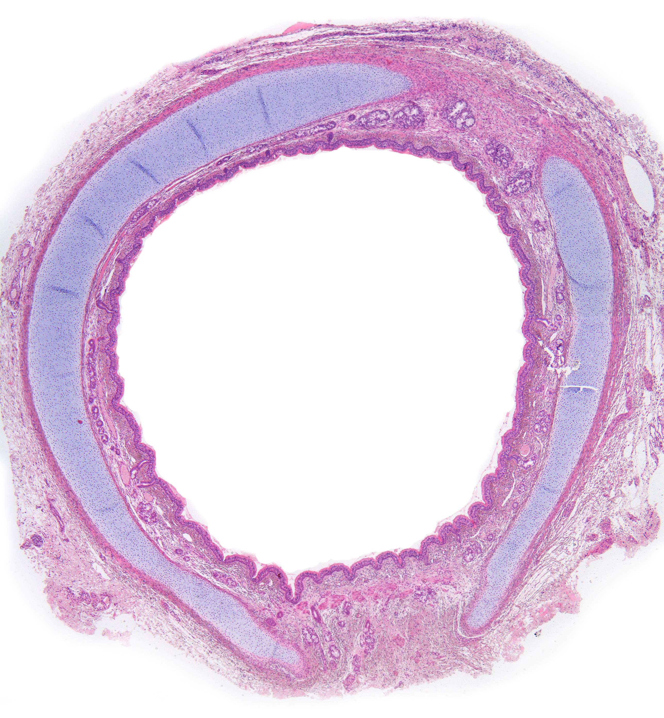
The windpipe (trachea) is a tubular organ made up of about 20 U-shaped hoops of hyaline cartilage with an opening at the back where you find smooth muscle that stiffens the wall.
This image shows a cross section of the trachea. The trachea is composed of several layers, including the mucosa, submucosa, tracheal cartilage, and adventitia. The mucosa contains a layer of ciliated pseudostratified columnar epithelium and goblet cells, while the submucosa contains seromucous glands. The tracheal cartilage provides structural support, and the adventitia contains blood vessels, nerves, and lymphatic vessels.
The opening in the cartilage at the top right is a cutting artefact.