
Vein and artery histology (40X)
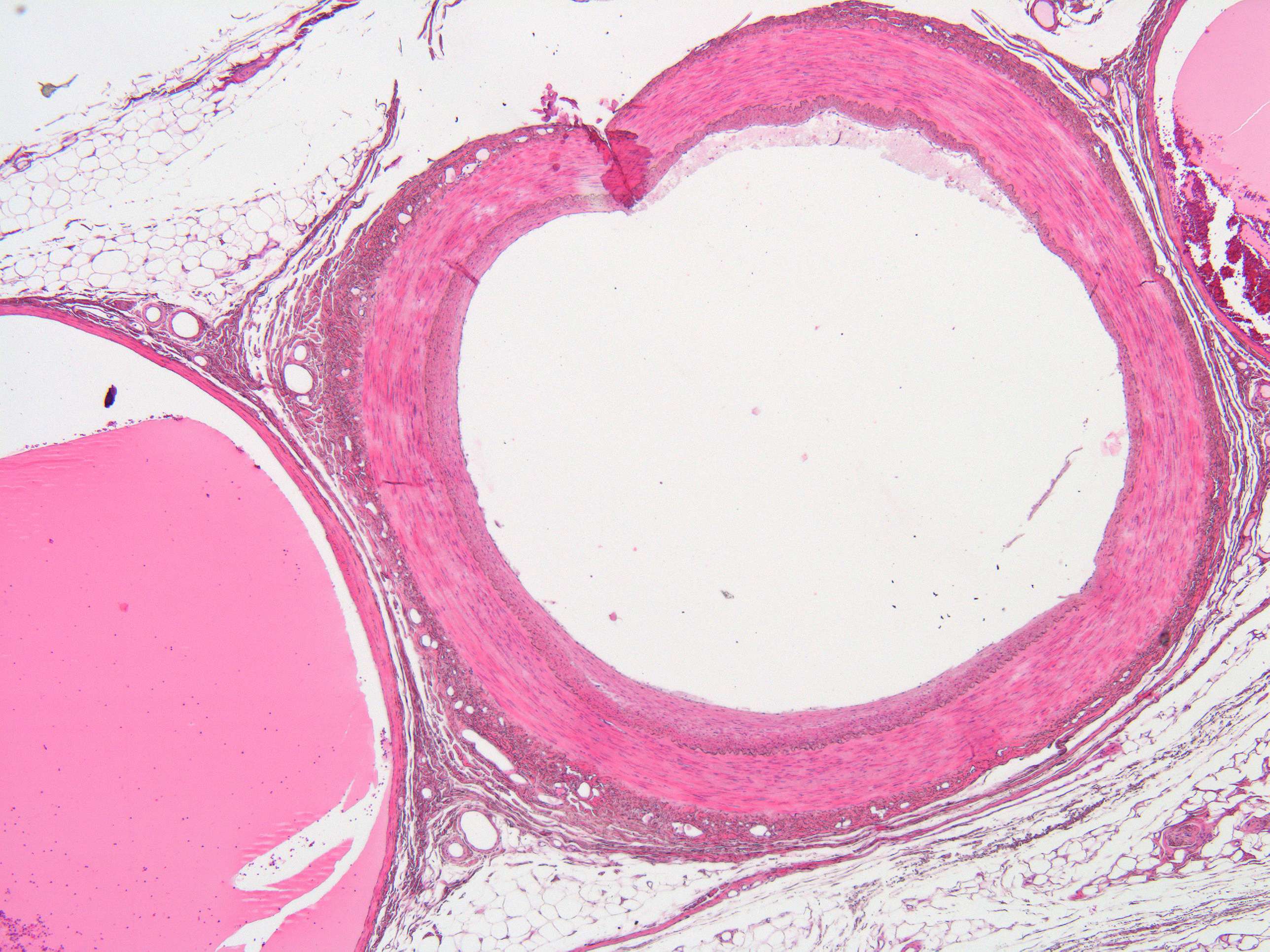
Ok, so you identified the artery. Well done! As you can see, the ertery has a much thicker wall than the vein. This is amongst other reasons that it has to support a much higher pressure from within than the vein. Arteries carry blood that is oxygenated after it has been pumped from the heart. Arteries consists of three tunics or layers: Tunica media, intima, and externa (adventitia).
Systemic arteries can be subdivided into two types—muscular and elastic—according to the relative compositions of elastic and muscle tissue in their tunica media as well as their size and the makeup of the internal and external elastic lamina. The larger arteries (>10 mm diameter) are generally elastic and the smaller ones (0.1–10 mm) tend to be muscular like the one you see here.
Veins have less smooth muscle, and connective tissue, and wider internal diameters than arteries. Because of their thinner walls and wider lumens they are able to expand and hold more blood.
Veins have a similar three-layered structure to arteries. The layers known as tunicas have a concentric arrangement that forms the wall of the vessel. The outer layer, is a thick layer of connective tissue called the tunica externa or adventitia; this layer is absent in the post-capillary venules. The middle layer, consists of bands of smooth muscle and is known as the tunica media. The inner layer, is a thin lining of endothelium known as the tunica intima. The tunica media in the veins is much thinner than that in the arteries as the veins are not subject to the high systolic pressures that the arteries are.