

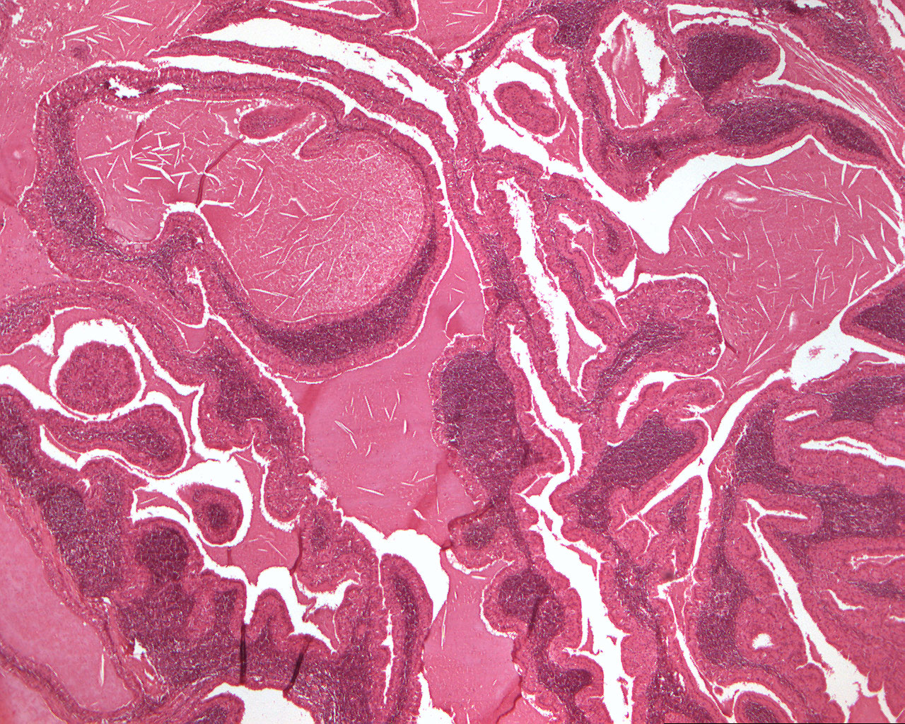
Papillary cystadenoma lymphomatosum (Warthin's tumor) (40X)
Clinical details: A 57-year-old man presented with a superficial, tumor-like bulge in the parotid region. He had noticed the process for approx. 2 years.
Clinical diagnosis: Tumor of the parotid region.
Microscopic examination: One finds numerous lumina (cysts) with papillomatous projections lined by a multi-rowed columnar epithelium with cells resembling oncocytes (oncos(G) = mass, kylos(G) = cell; large, finely granular, highly eosinophilic cells). The lumen of the cysts, which can be quite large, contains an eosinophilic substance where a number of leukocytes can be seen, as well as slit-like cavities due to cholesterol crystals. Subepithelially, one finds abundant lymphocytes, partly with the presence of regular lymphatic tissue (B lymphocytes) with germinal centers.