
The neural tube (40X)
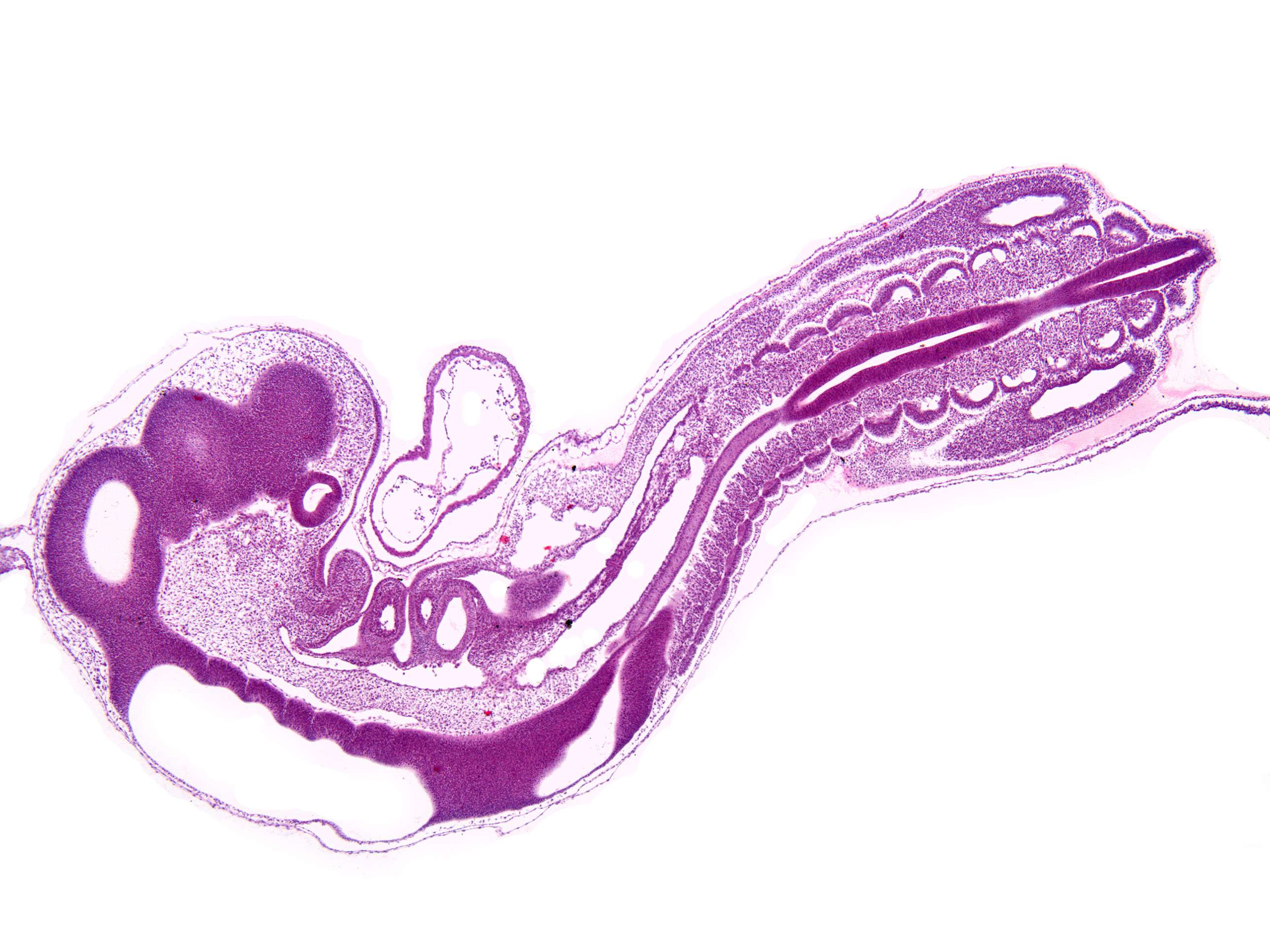
This is a longitudonal section through a chicken fetus. During the fixation, some parts of the fetus has rotated perpendicular to its longitudinal axis, so that the section through the head is more or less a sagittal sction, while the posterior (caudal) part is more or less a frontal section (note somites symmetrically on each side of the neural tube that would not be visible in a purely saggital section).
The section shows the neural tube shortly after it has closed and the development of brain vesicles has begun. In addition, cardiac structures and somites are seen.
It is recomended to read about the early development of the central nervous system before studying this section. try to identify the:
- Mesencephalon
- Proencephalon
- Neural tube
- Rhombencephalon
- Somites