
The otic vesicle of an embryo (40X)
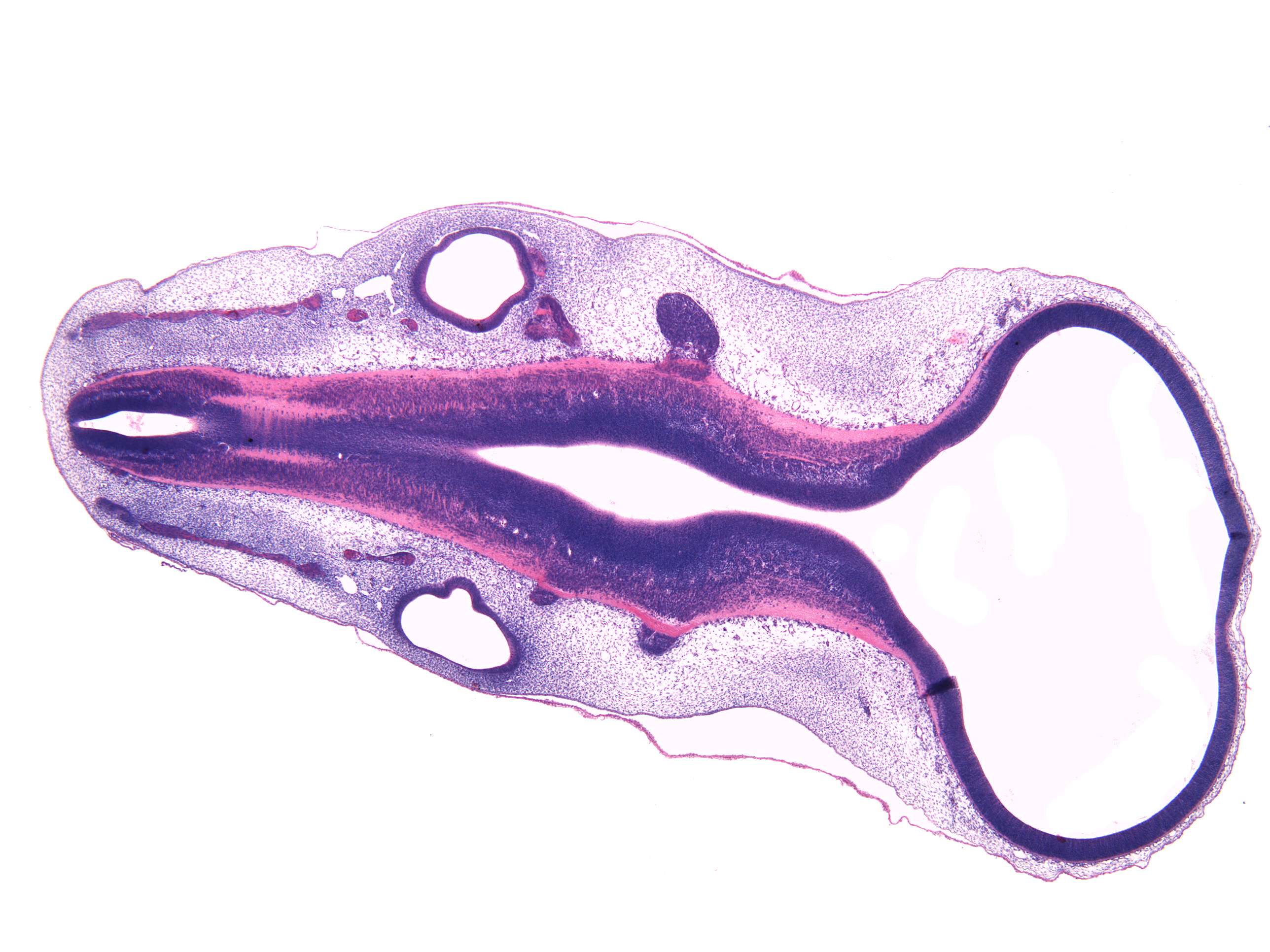
The section shows development of the inner ear (otic vesicle), and early development of the brainstem and cranial nerves. The otic vesicle is formed by detachment from the ectoderm, and develops into the cochlea, archways and sacculus and utriculus. The eardrum loses its connection with the surface early on.
The anatomical term myotome which describes the muscles served by a spinal nerve root, is also used in embryology to describe that part of the somite which develops into the muscles as seen here.