
Palpebrae (40X)
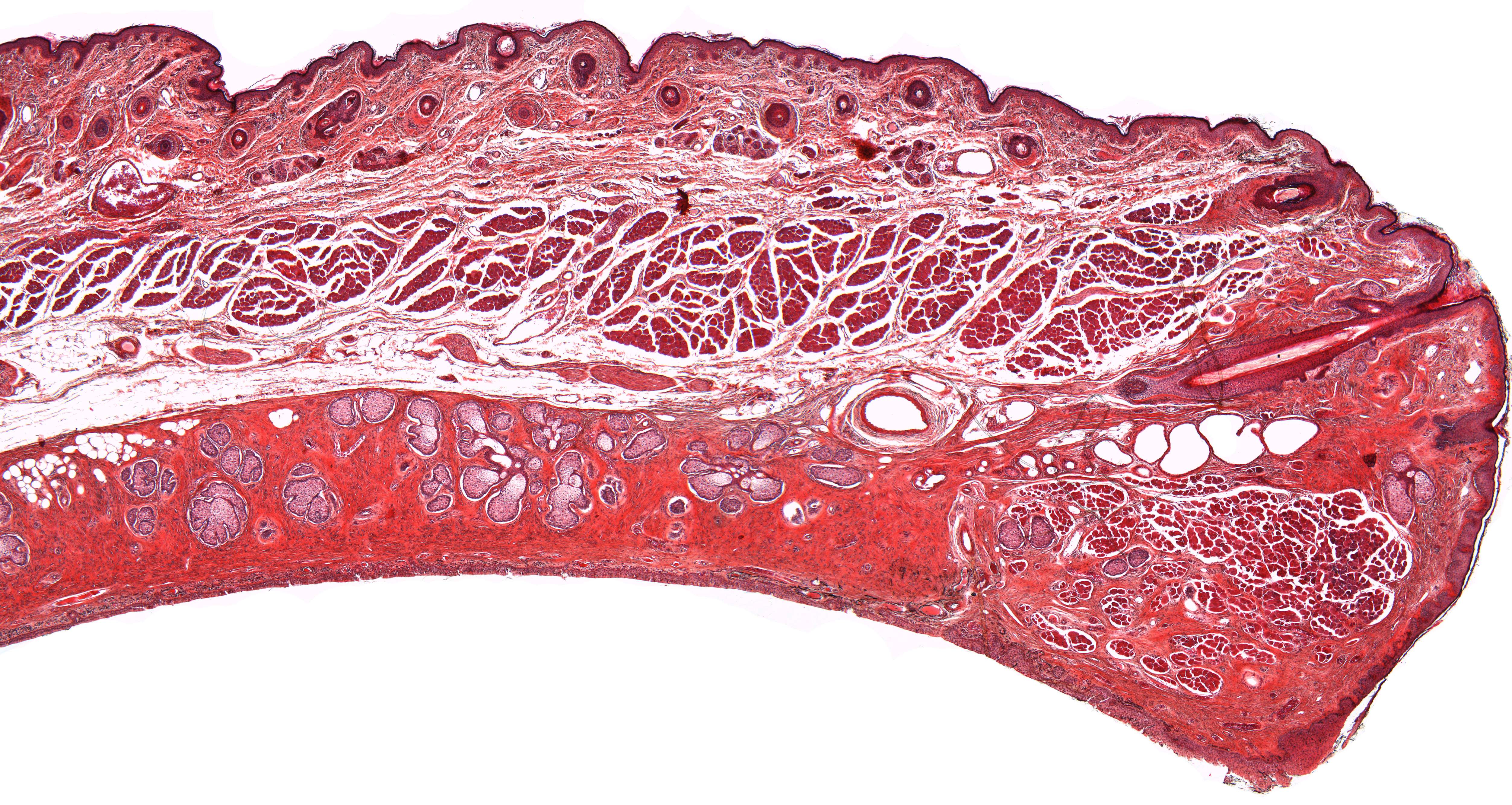
This images shows a few more details of the lower rim of the palpebra (lower end facing to the far right). Samll hair follicles are seen below the squamous epithelium covering the outer surface of the palpebra. Compare the size of the hair follicles to the one that is seen at the base of the cilium (eye lash).
The tissue facing the eyeball as a whole is called the tarsal plate. The epithelium covering the tarsal plate facing the eyeball is of the low, stratified columnar type. Below the epithelium and within the tarsal plate, there are tarsal glands. These glands are sebaceous glands that produce meibum, an oily substance that prevents evaporation of the eye's tear film.
A cross sectrion of the orbicularis oculi muscle can be seen at the center thouroughout the eye lid