
Sinusoids of the human liver (400X)
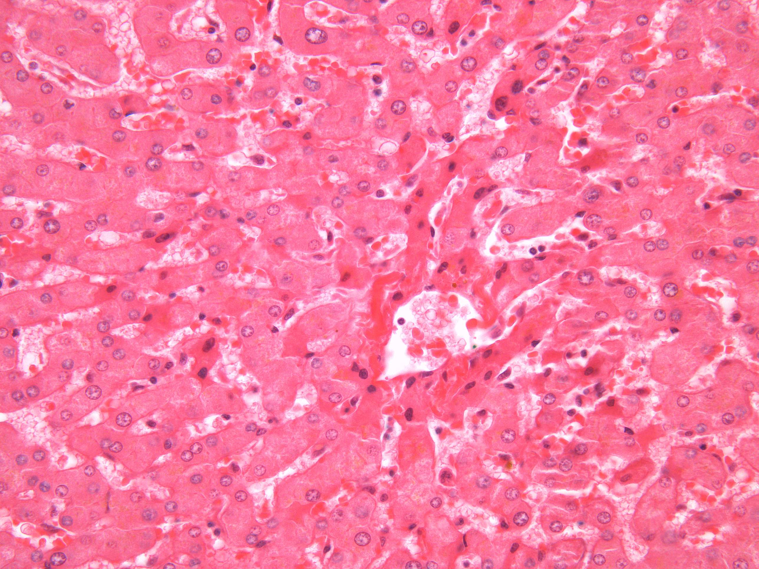
Sinusoids are large wide capillaries, with a diameter of 30-40 μm or more. In the liver, the sinusoids form the boundary between the lamellae of liver cells. They are therefore not cylindrical, but adapt to the size of the space between two neighboring liver cell lamellae.
Within the sinusoids of the liver one naturally finds the blood cells (red and white blood cells). In addition, endothelial cells and Kupfer cells which are stationary macrophages that are situated within the wall of the sinusoids. In a H+E section like this, one can easily point out the endothelial cells and the blood cells, but it is not so easy to spot the Kupfer cells (the cells have a dark round nucleus and next to no cytoplasm).