
The ischiadic nerve (200X)
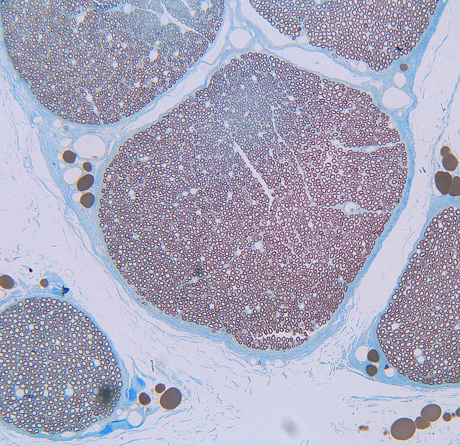
The image shows a cross section of the ischiadic nerve from a cat.
The preparation is particularly well suited to identify large and small myelinated axons, the perineurium and capillaries inside the fascicles.
The adipose / fat cells are colored light brown in this section and are seen next to the perineurium. The perineurium is coloured blue.
Try to identify;
- adipose cells (fat cells)
- fascicles