
Pylorus and duodenum
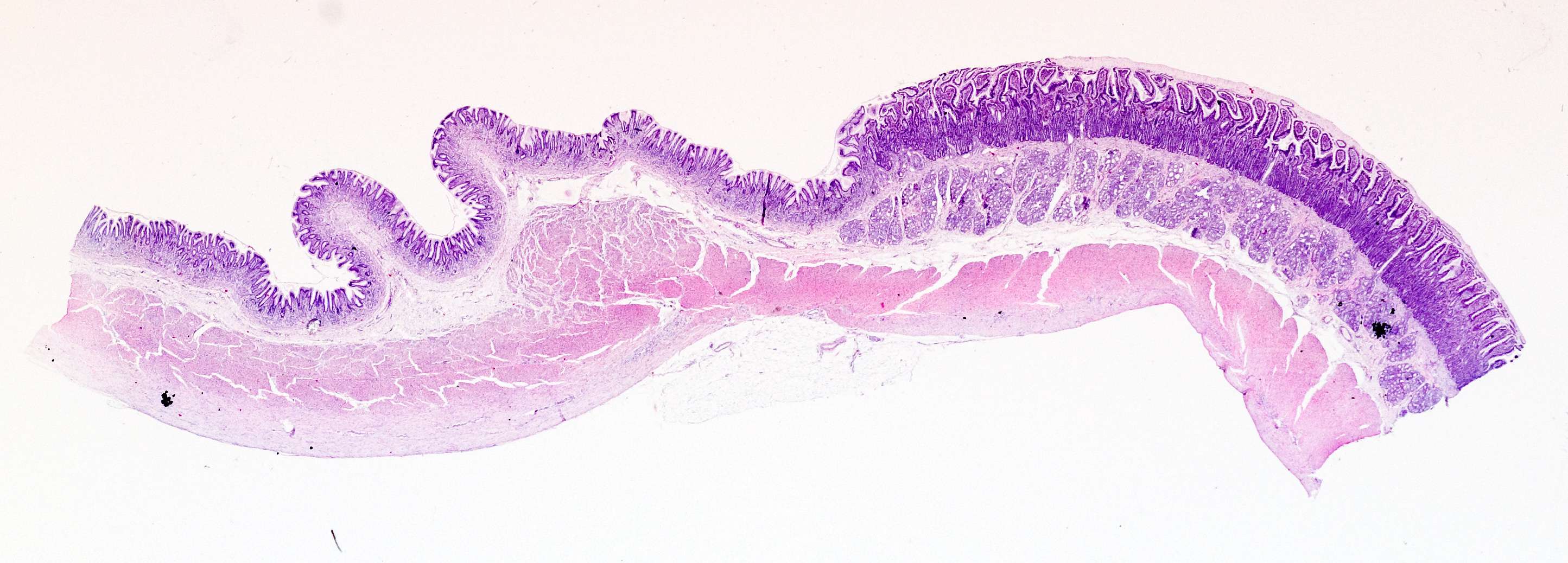
In this image you can see the transitional zone between the mucosa in the pylorus and in the duodenum.
On the duodenal side, i.e. in the small intestine, villi intestinales and scattered goblet cells are seen throughout the columnar epithelium. Villi appear partly as oblong structures claed with columnar epithelium connected to the underlying layer, partly as isolated islands made with columnar epithelium. Also characteristic of the tunica mucosa of the small intestine is the submucous glands in duodenum (Brunner's glands). The Brunner's glands (or duodenal glands) produce HCO3-which helps neutralize the acidic chymus from the ventricle.