
Colon histology
The colon is the largest portion of the large intestine. Most sources define the large intestine as the combination of the cecum, colon, rectum, and anal canal.
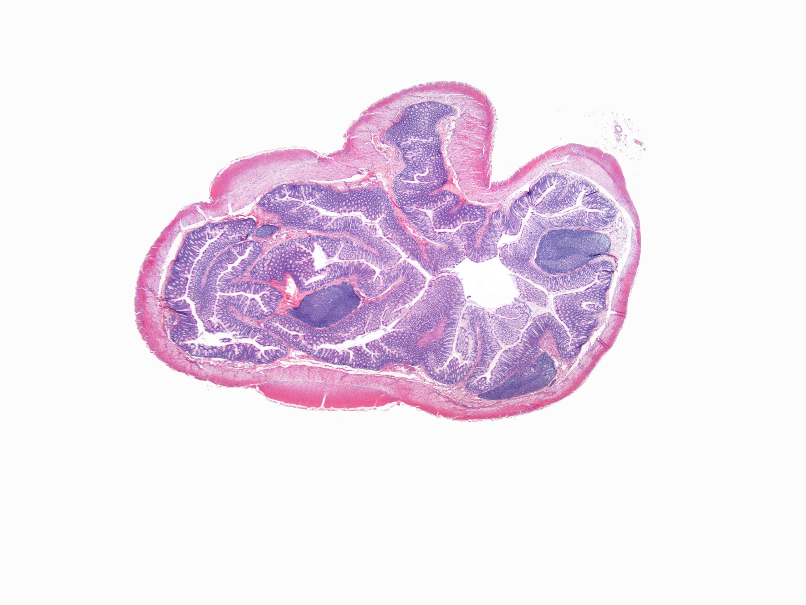
This image is a cross section of the colon. Microscopically, the colon crypts are shaped like microscopic thick walled test tubes with a central hole down the length of the tube (the crypt lumen). Four tissue sections are shown here, two cut across the long axes of the crypts and two cut parallel to the long axes.
Note in this section:
- Intestinal glands (crypts of Lieberkühn)
- Tunica muscularis with circular and longitudinal muscle cells