
Spermatogonium and Leydig cell
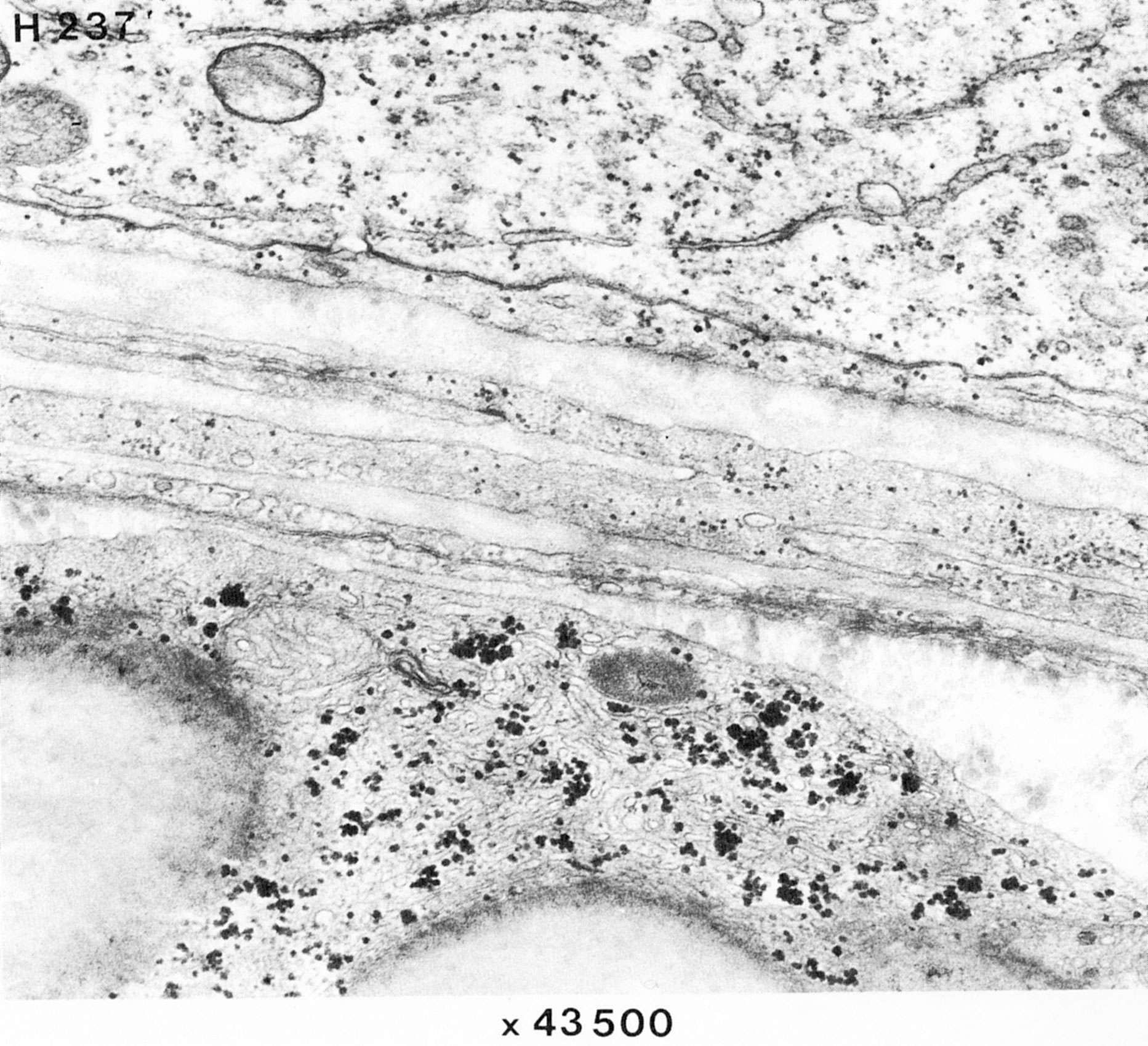
This picture shows the outemost layer of a semineferous tubule, consisting mainly of myoepithelial cells (and some collagen fibrils in cross section). Note the cell junctions among the myoepithelial cells. The Leydig cell below shows abundant smooth endoplasmic reticulum, which is typical of steroid-producing cells.