
Placenta (3 months) (100X)
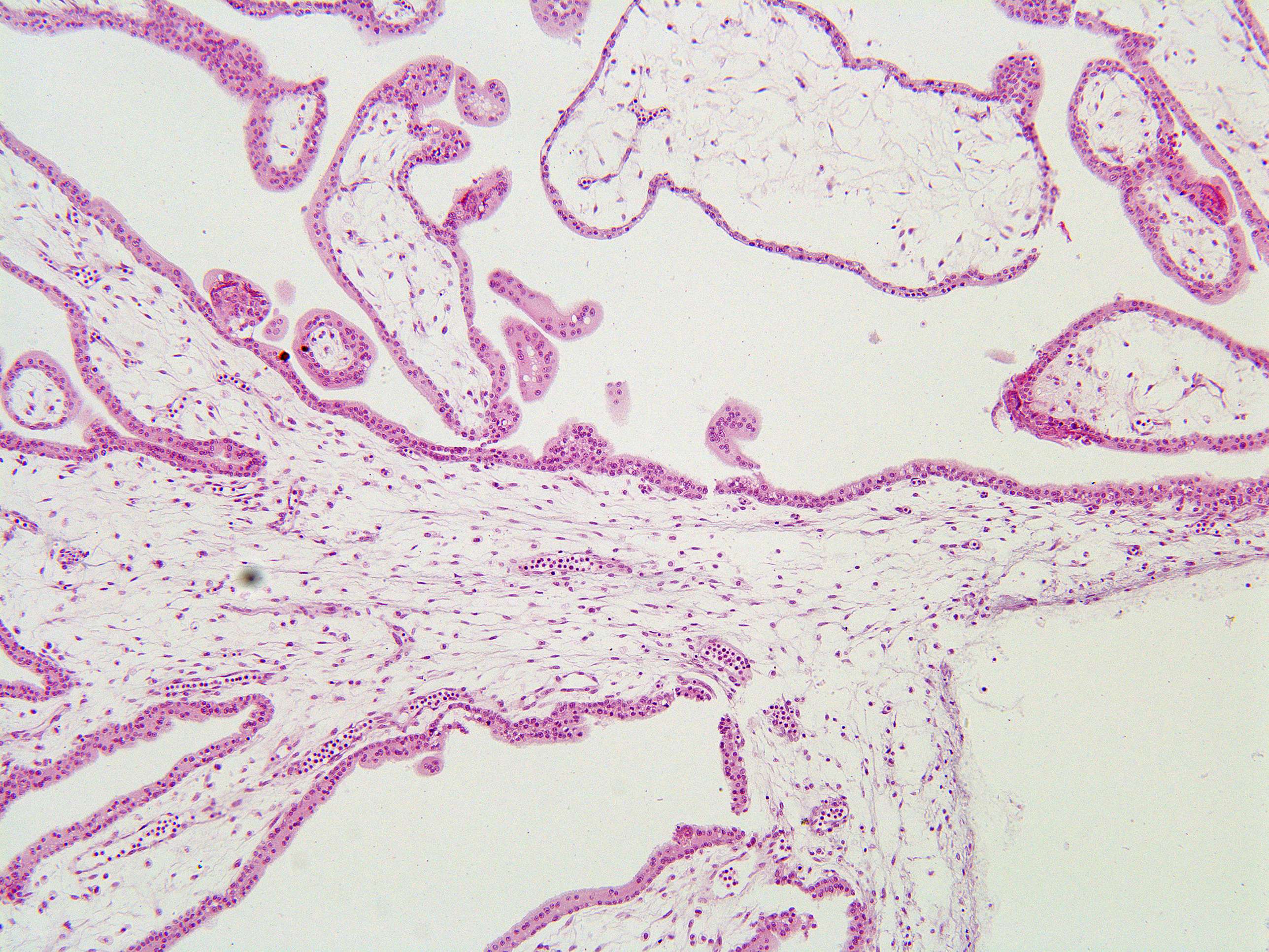
The chorionic plate and the villi. In the extraembryonic mesoderm, some small blood vessels are seen. Note that the erythrocytes are nucleated. The trophoblast is lining the villi and the chorionic plate. The cytotrophoblast layer, closest to the extraembryonic mesoderm, can easily be distinguished from the syncytiotrophoblast layer.