
Vaginal smear (400X)
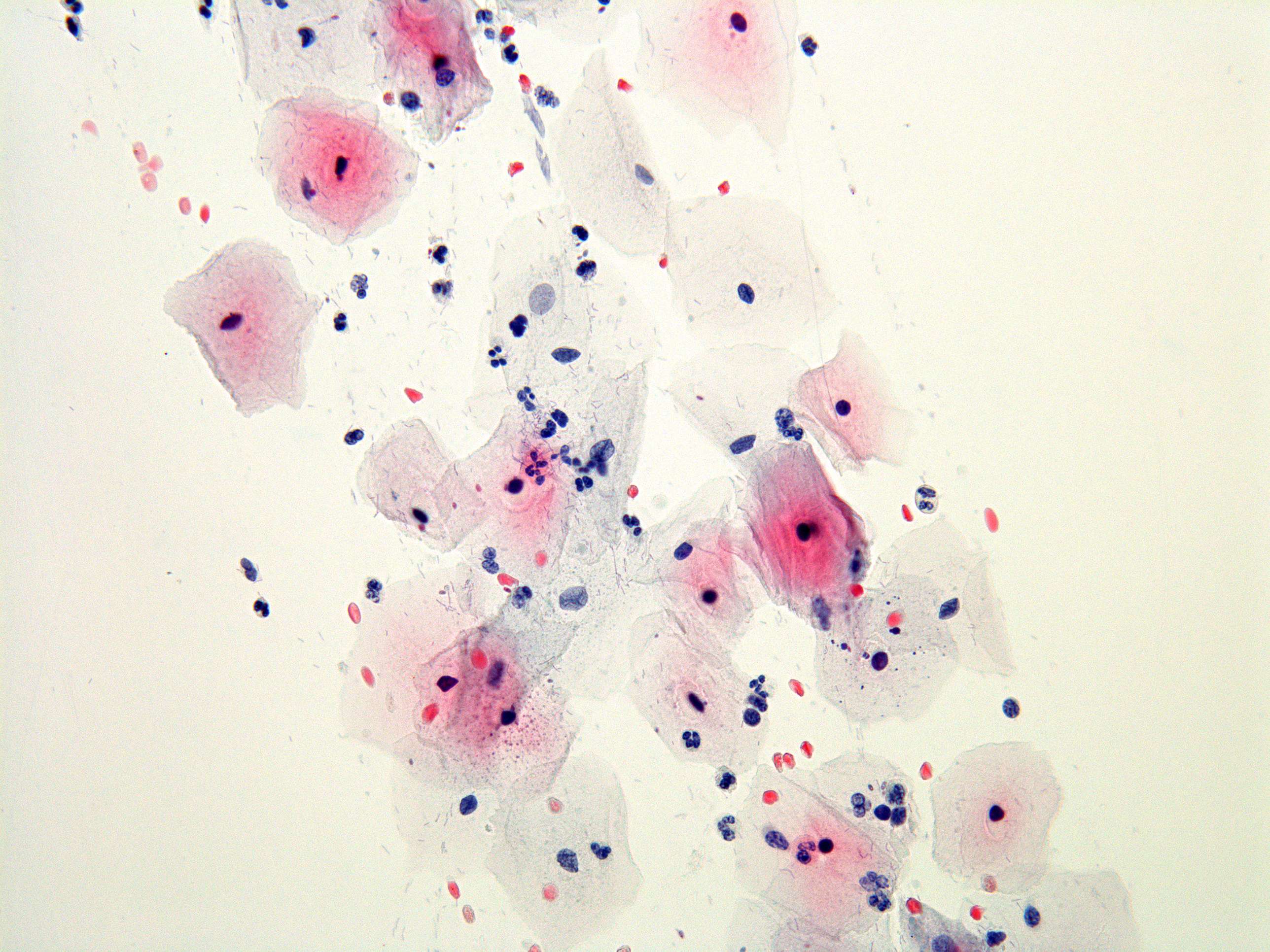
You see squamous epithelial cells (squamous cells) which are normally lining the vaginal wall. You also se granulocytes with their characteristically dark and lobular nuclei.
You see squamous epithelial cells (squamous cells) which are normally lining the vaginal wall. You also se granulocytes with their characteristically dark and lobular nuclei.